Eichelberger Dominique, Calabrese Pasquale, Meyer Antonia, Chaturvedi Menorca, Hatz Florian, Fuhr Peter, Gschwandtner Ute
Division of Molecular and Cognitive Neuroscience, Neuropsychology and Behavioural Neurology Unit, University of Basel, Basel, Switzerland.
Department of Neurology, Hospital of the University of Basel, Petersgraben 4, 4031 Basel, Switzerland.
Parkinsons Dis. 2017;2017:3659784. doi: 10.1155/2017/3659784. Epub 2017 Feb 28.
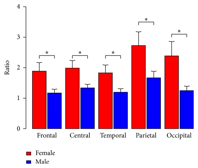
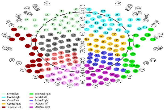
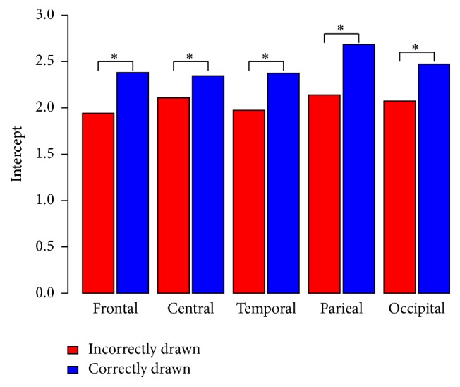
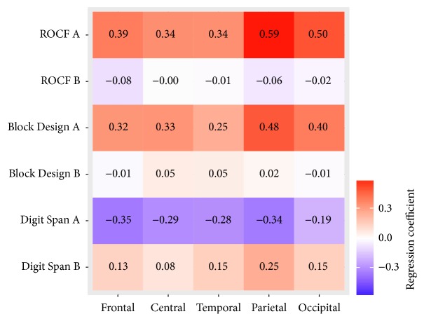
Visuospatial dysfunction is among the first cognitive symptoms in Parkinson's disease (PD) and is often predictive for PD-dementia. Furthermore, cognitive status in PD-patients correlates with quantitative EEG. This cross-sectional study aimed to investigate the correlation between EEG slowing and visuospatial ability in nondemented PD-patients. Fifty-seven nondemented PD-patients (17 females/40 males) were evaluated with a comprehensive neuropsychological test battery and a high-resolution 256-channel EEG was recorded. A median split was performed for each cognitive test dividing the patients sample into either a normal or lower performance group. The electrodes were split into five areas: frontal, central, temporal, parietal, and occipital. A linear mixed effects model (LME) was used for correlational analyses and to control for confounding factors. Subsequently, for the lower performance, LME analysis showed a significant positive correlation between ROCF score and parietal alpha/theta ratio ( = .59, = .012) and occipital alpha/theta ratio ( = 0.50, = .030). No correlations were found in the group of patients with normal visuospatial abilities. We conclude that a reduction of the parietal alpha/theta ratio is related to visuospatial impairments in PD-patients. These findings indicate that visuospatial impairment in PD-patients could be influenced by parietal dysfunction.
视觉空间功能障碍是帕金森病(PD)最早出现的认知症状之一,且常常是PD痴呆的预测指标。此外,PD患者的认知状态与定量脑电图相关。这项横断面研究旨在调查非痴呆PD患者脑电图减慢与视觉空间能力之间的相关性。对57名非痴呆PD患者(17名女性/40名男性)进行了全面的神经心理测试,并记录了高分辨率256通道脑电图。对每项认知测试进行中位数分割,将患者样本分为表现正常或较低的组。电极被分为五个区域:额叶、中央、颞叶、顶叶和枕叶。采用线性混合效应模型(LME)进行相关性分析并控制混杂因素。随后,对于表现较低的情况,LME分析显示复制 Rey 复杂图形测验(ROCF)得分与顶叶α/θ比值(r = 0.59,p = 0.012)和枕叶α/θ比值(r = 0.50,p = 0.030)之间存在显著正相关。在视觉空间能力正常的患者组中未发现相关性。我们得出结论,顶叶α/θ比值降低与PD患者的视觉空间障碍有关。这些发现表明,PD患者的视觉空间障碍可能受顶叶功能障碍影响。