Liang Jinying, Zhang Xinxin, Miao Yunqiu, Li Juan, Gan Yong
Department of Pharmaceutics, China Pharmaceutical University, Nanjing, People's Republic of China; Shanghai Institute of Materia Medica, Chinese Academy of Sciences, Shanghai, People's Republic of China; School of Pharmacy, Xinxiang Medical University, Xinxiang, People's Republic of China.
Shanghai Institute of Materia Medica, Chinese Academy of Sciences, Shanghai, People's Republic of China.
Int J Nanomedicine. 2017 Mar 14;12:2033-2044. doi: 10.2147/IJN.S128525. eCollection 2017.
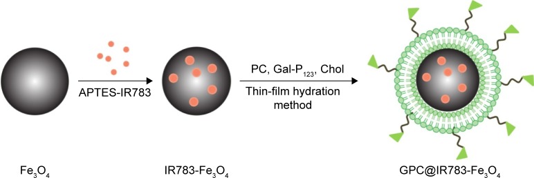
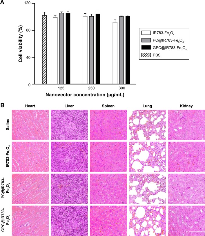
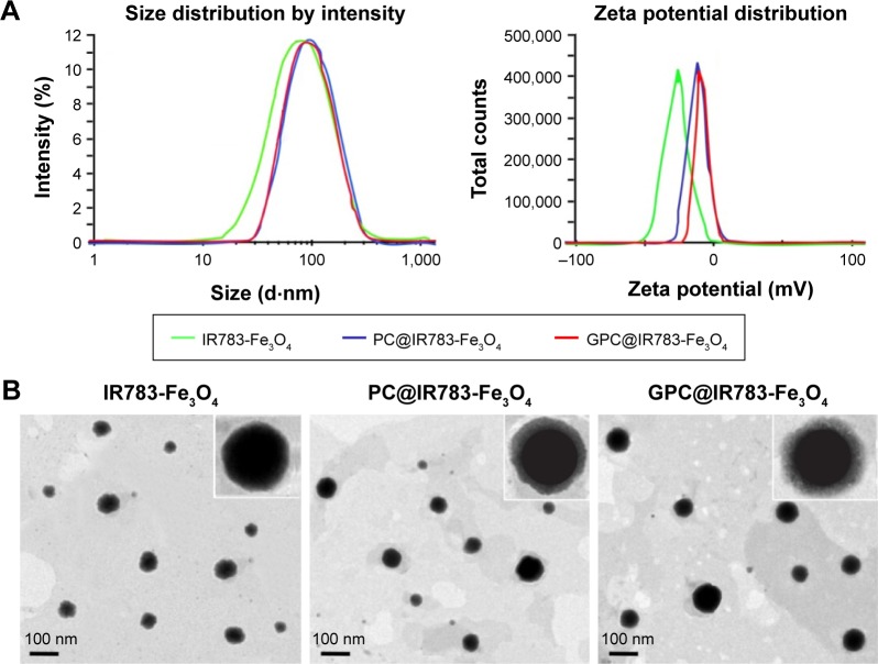
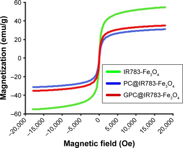
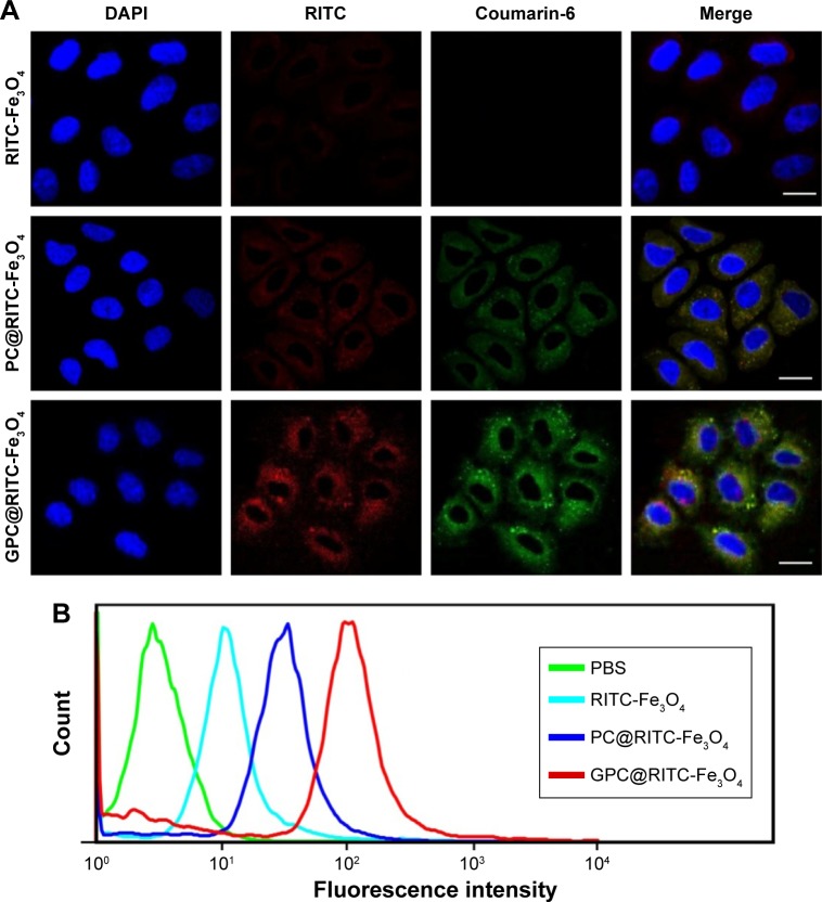
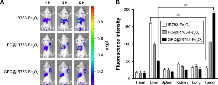
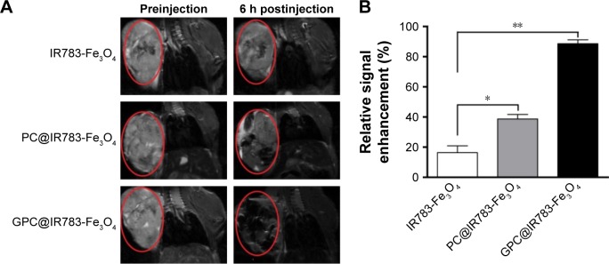
The development of noninvasive imaging techniques for the accurate diagnosis of progressive hepatocellular carcinoma (HCC) is of great clinical significance and has always been desired. Herein, a hepatocellular carcinoma cell-targeting fluorescent magnetic nanoparticle (NP) was obtained by conjugating near-infrared fluorescence to the surface of FeO (NIRF-FeO) NPs, followed by coating the lipids consisting of tumoral hepatocytes-targeting polymer (Gal-P). This magnetic NP (GPC@NIRF-FeO) with superparamagnetic behavior showed high stability and safety in physiological conditions. In addition, GPC@NIRF-FeO achieved more specific uptake of human liver cancer cells than free FeO NPs. Importantly, with superpara-magnetic iron oxide and strong NIR absorbance, GPC@NIRF-FeO NPs demonstrate prominent tumor-contrasted imaging performance both on fluorescent and T-weighted magnetic resonance (MR) imaging modalities in a living body. The relative MR signal enhancement of GPC@NIRF-FeO NPs achieved 5.4-fold improvement compared with NIR-FeO NPs. Therefore, GPC@ NIRF-FeO NPs may be potentially used as a candidate for dual-modal imaging of tumors with information covalidated and directly compared by combining fluorescence and MR imaging.
开发用于准确诊断进展期肝细胞癌(HCC)的非侵入性成像技术具有重大临床意义,一直是人们所期望的。在此,通过将近红外荧光缀合到FeO(NIRF-FeO)纳米颗粒表面,然后包覆由靶向肿瘤肝细胞的聚合物(Gal-P)组成的脂质,获得了一种靶向肝细胞癌细胞的荧光磁性纳米颗粒(NP)。这种具有超顺磁性的磁性NP(GPC@NIRF-FeO)在生理条件下表现出高稳定性和安全性。此外,GPC@NIRF-FeO对人肝癌细胞的摄取比游离FeO纳米颗粒更具特异性。重要的是,凭借超顺磁性氧化铁和强近红外吸收,GPC@NIRF-FeO纳米颗粒在活体的荧光和T加权磁共振(MR)成像模态上均表现出突出的肿瘤对比成像性能。与NIR-FeO纳米颗粒相比,GPC@NIRF-FeO纳米颗粒的相对MR信号增强提高了5.4倍。因此,GPC@NIRF-FeO纳米颗粒可能有潜力作为一种用于肿瘤双模态成像的候选物,通过结合荧光和MR成像来验证和直接比较信息。