Li Jun, Zhao Lei, Urabe Go, Fu Yingmei, Guo Lian-Wang
Department of Ophthalmology, The First Hospital of China Medical University, Shenyang, China; Department of Ophthalmology, The 3rd People's Hospital of Dalian, Dalian, China; Department of Surgery, 5151 Wisconsin Institute for Medical Research, University of Wisconsin-Madison, Madison, WI.
Department of Surgery, 5151 Wisconsin Institute for Medical Research, University of Wisconsin-Madison, Madison, WI.
Mol Vis. 2017 Mar 21;23:149-159. eCollection 2017.
The bromo and extraterminal (BET) epigenetic "reader" family is becoming an appealing new therapeutic target for several common diseases, yet little is known of its role in retinal neurodegeneration. We explored the potential of BET inhibition in the protection of retinal ganglion cells (RGCs).
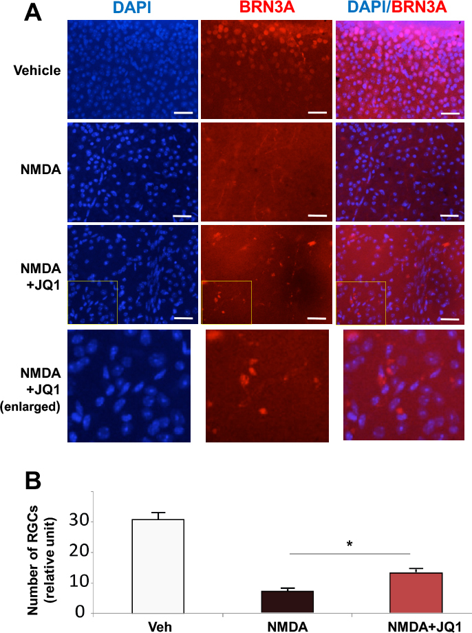
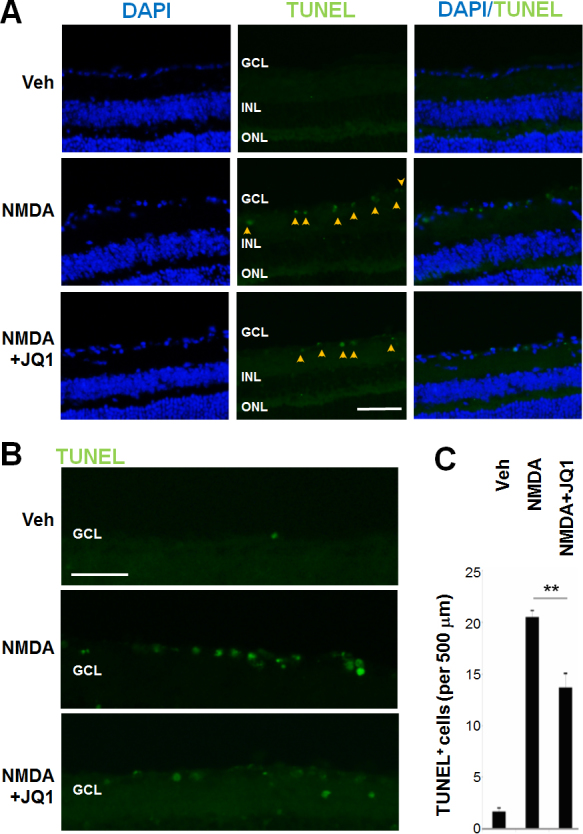
To test the therapeutic effect of JQ1, an inhibitor highly selective for the BET family of proteins, we used an acute RGC damage model induced by N-methyl-D-aspartic acid (NMDA) excitotoxicity. Adult C57BL/6 mice received an intravitreal injection of NMDA with (or without) JQ1 in one eye and vehicle control in the contralateral eye; RGC loss was assessed on retinal sections and whole mounts. Gene expression and apoptosis were analyzed by quantitative real time (RT)-PCR and terminal deoxynucleotidyl transferase dUTP nick end labeling (TUNEL), respectively. For counting RGCs, immunostaining of the marker protein BRN3A was performed on whole mounts.
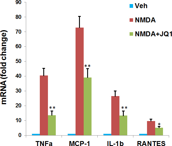
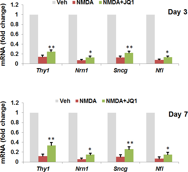
NMDA treatment eliminated RGCs (day 7 and day 14 post injection) and diminished the expression (mRNAs) of RGC-selective genes, including , and (day 3 and day 7). In contrast, co-injection with JQ1 maintained the number and gene expression of RGCs at ~2 fold of the control (NMDA only, no JQ1), and it decreased NMDA-induced TUNEL-positive cells in the RGC layer by 35%. While NMDA treatment dramatically upregulated mRNAs of inflammatory cytokines (TNFα, IL-1β, MCP-1, RANTES) in retinal homogenates, co-injection with JQ1 suppressed their upregulation by ~50%.
Intravitreal injection of a BET inhibitor (JQ1) ameliorates NMDA-induced RGC death, revealing the RGC-protective potential of pharmacological blockage of the BET family. This new strategy of epigenetic intervention may be extended to other retinal degenerative conditions.
溴结构域与额外末端(BET)表观遗传“读取器”家族正成为几种常见疾病有吸引力的新治疗靶点,但其在视网膜神经退行性变中的作用知之甚少。我们探讨了BET抑制在保护视网膜神经节细胞(RGC)方面的潜力。
为了测试对BET蛋白家族具有高度选择性的抑制剂JQ1的治疗效果,我们使用了由N-甲基-D-天冬氨酸(NMDA)兴奋性毒性诱导的急性RGC损伤模型。成年C57BL/6小鼠一只眼睛玻璃体内注射NMDA加(或不加)JQ1,对侧眼睛注射溶媒对照;在视网膜切片和整装片上评估RGC损失。分别通过定量实时(RT)-PCR和末端脱氧核苷酸转移酶dUTP缺口末端标记(TUNEL)分析基因表达和细胞凋亡。为了计数RGC,在整装片上对标记蛋白BRN3A进行免疫染色。
NMDA处理导致RGC消失(注射后第7天和第14天),并降低了RGC选择性基因的表达(mRNA),包括[此处基因名称缺失]和[此处基因名称缺失](第3天和第7天)。相比之下,与JQ1共同注射使RGC的数量和基因表达维持在对照(仅NMDA,无JQ1)的约2倍,并使RGC层中NMDA诱导的TUNEL阳性细胞减少35%。虽然NMDA处理显著上调了视网膜匀浆中炎性细胞因子(TNFα、IL-1β、MCP-1、RANTES)的mRNA,但与JQ1共同注射将它们的上调抑制了约50%。
玻璃体内注射BET抑制剂(JQ1)可改善NMDA诱导的RGC死亡,揭示了BET家族药物阻断的RGC保护潜力。这种表观遗传干预的新策略可能扩展到其他视网膜退行性疾病。