Daniels Brian P, Snyder Annelise G, Olsen Tayla M, Orozco Susana, Oguin Thomas H, Tait Stephen W G, Martinez Jennifer, Gale Michael, Loo Yueh-Ming, Oberst Andrew
Department of Immunology, University of Washington, Seattle, WA 98109, USA.
Molecular and Cellular Biology Program, University of Washington, Seattle, WA 98109, USA.
Cell. 2017 Apr 6;169(2):301-313.e11. doi: 10.1016/j.cell.2017.03.011. Epub 2017 Mar 30.
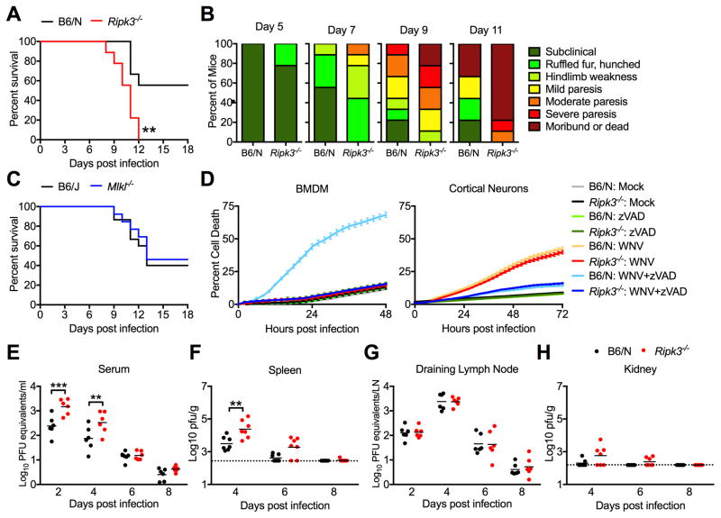
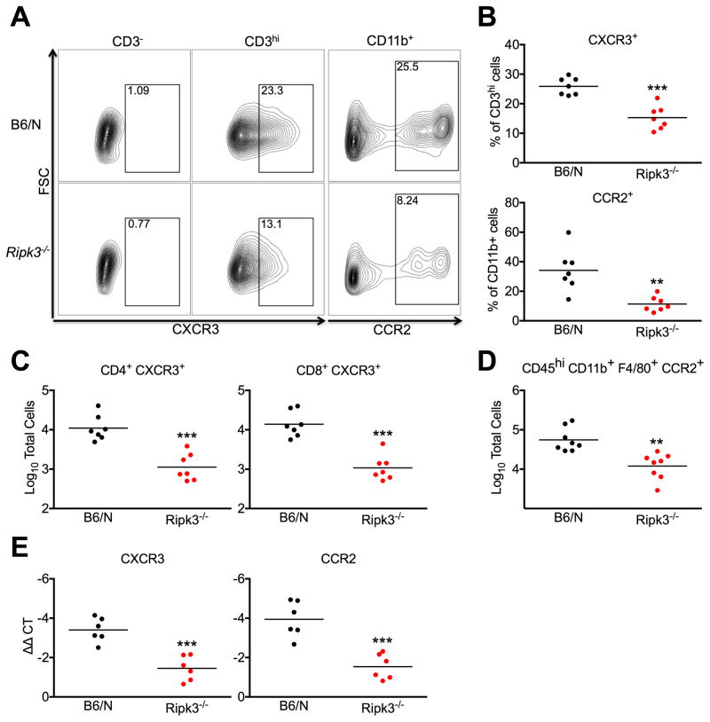
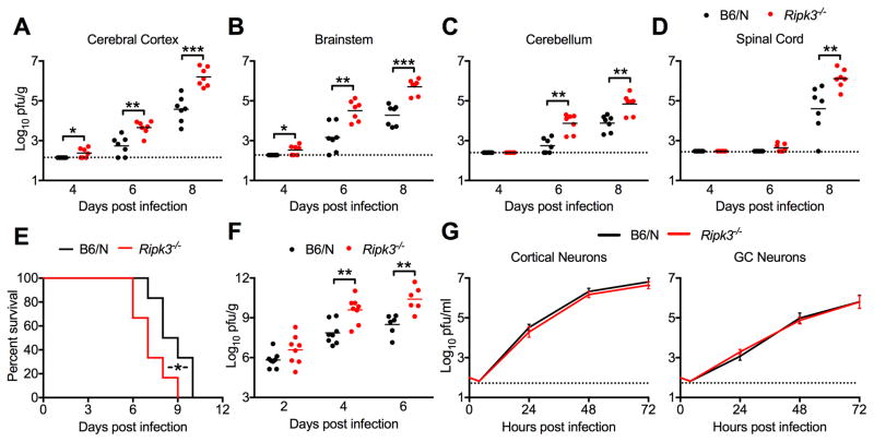
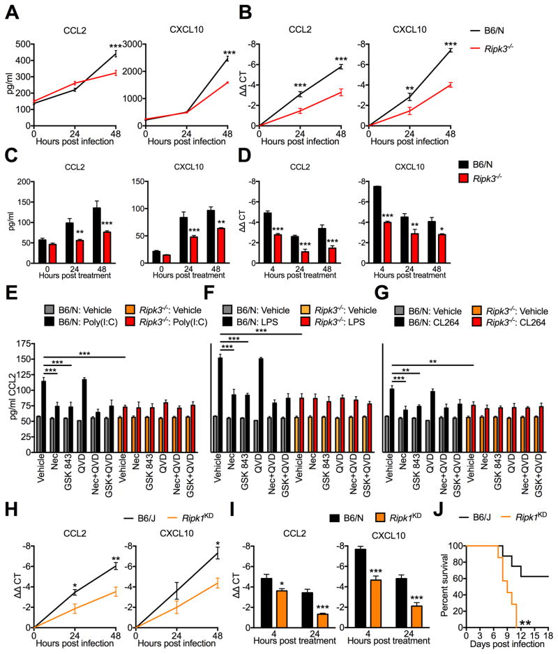
Receptor-interacting protein kinase-3 (RIPK3) is an activator of necroptotic cell death, but recent work has implicated additional roles for RIPK3 in inflammatory signaling independent of cell death. However, while necroptosis has been shown to contribute to antiviral immunity, death-independent roles for RIPK3 in host defense have not been demonstrated. Using a mouse model of West Nile virus (WNV) encephalitis, we show that RIPK3 restricts WNV pathogenesis independently of cell death. Ripk3 mice exhibited enhanced mortality compared to wild-type (WT) controls, while mice lacking the necroptotic effector MLKL, or both MLKL and caspase-8, were unaffected. The enhanced susceptibility of Ripk3 mice arose from suppressed neuronal chemokine expression and decreased central nervous system (CNS) recruitment of T lymphocytes and inflammatory myeloid cells, while peripheral immunity remained intact. These data identify pleiotropic functions for RIPK3 in the restriction of viral pathogenesis and implicate RIPK3 as a key coordinator of immune responses within the CNS.
受体相互作用蛋白激酶3(RIPK3)是坏死性细胞死亡的激活剂,但最近的研究表明,RIPK3在独立于细胞死亡的炎症信号传导中还有其他作用。然而,虽然坏死性凋亡已被证明有助于抗病毒免疫,但RIPK3在宿主防御中与细胞死亡无关的作用尚未得到证实。利用西尼罗河病毒(WNV)脑炎小鼠模型,我们发现RIPK3独立于细胞死亡限制WNV发病机制。与野生型(WT)对照相比,Ripk3小鼠死亡率更高,而缺乏坏死性凋亡效应蛋白MLKL或同时缺乏MLKL和半胱天冬酶-8的小鼠则未受影响。Ripk3小鼠易感性增加是由于神经元趋化因子表达受抑制,以及T淋巴细胞和炎性髓细胞向中枢神经系统(CNS)募集减少,而外周免疫保持完整。这些数据确定了RIPK3在限制病毒发病机制中的多效性功能,并表明RIPK3是CNS内免疫反应的关键协调者。