Ramezani Mehdi, Salehnia Mojdeh, Jafarabadi Mina
Department of Anatomical Sciences, Medical Sciences Faculty, Tarbiat Modares University, Tehran, Iran.
Reproductive Health Research Center, Tehran University of Medical Sciences, Tehran, Iran.
J Reprod Infertil. 2017 Jan-Mar;18(1):162-171.
The aim of the present study was to evaluate the effectiveness of human ovarian vitrification protocol followed with culture at the morphological and molecular levels.
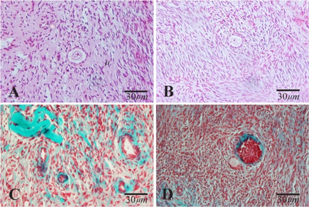
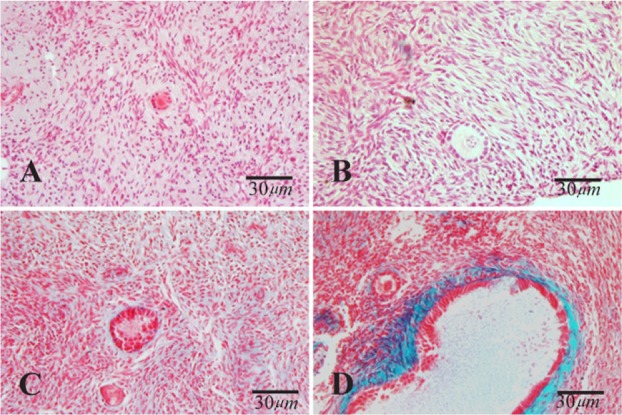
Ovarian tissues were obtained from 10 normal transsexual women and cut into small pieces and were divided into non-vitrified and vitrified groups and some of the tissues fragments in both groups were randomly cultured for two weeks. The morphological study using hematoxylin and eosin and Masson's trichrome staining was done. The analysis of mean follicular density, 17-β estradiol (E2) and anti mullerian hormone (AMH), and real-time RT-PCR was down for the evaluation of expression of genes related to folliculogenesis. Data were compared by paired-samples and independent-samples T test. Values of p<0.05 were considered statistically significant.
The proportion of normal follicles did not show significant difference between vitrified and non-vitrified groups before and after culture but these rates and the mean follicle density significantly decreased in both cultured tissues (p<0.05). The expression of genes was similar in vitrified and non-vitrified groups but in cultured tissues the expression of GDF9 and FSHR genes increased and the expression of FIGLA and KIT-L genes decreased (p<0.05). An increase in E2 and AMH concentration was observed after 14 days of culture in both groups.
In conclusion, the present study indicated that the follicular development and gene expression in vitrified ovarian tissue was not altered before and after culture, thus this method could be useful for fertility preservation; however, additional studies are needed to improve the culture condition.
本研究的目的是在形态学和分子水平上评估人类卵巢玻璃化方案及随后培养的有效性。
从10名正常变性女性获取卵巢组织,切成小块,分为未玻璃化组和玻璃化组,两组中的一些组织碎片随机培养两周。使用苏木精和伊红以及马松三色染色进行形态学研究。分析平均卵泡密度、17-β雌二醇(E2)和抗苗勒管激素(AMH),并进行实时逆转录聚合酶链反应以评估与卵泡发生相关基因的表达。数据通过配对样本和独立样本t检验进行比较。p<0.05的值被认为具有统计学意义。
培养前后,玻璃化组和未玻璃化组的正常卵泡比例无显著差异,但两组培养组织中的这些比率和平均卵泡密度均显著降低(p<0.05)。玻璃化组和未玻璃化组的基因表达相似,但在培养组织中,生长分化因子9(GDF9)和促卵泡激素受体(FSHR)基因的表达增加,而卵泡生成素样蛋白A(FIGLA)和干细胞因子配体(KIT-L)基因的表达降低(p<0.05)。两组培养14天后均观察到E2和AMH浓度增加。
总之,本研究表明,玻璃化卵巢组织在培养前后的卵泡发育和基因表达未发生改变,因此该方法可能有助于生育力保存;然而,需要进一步研究以改善培养条件。