Tarkin Jason M, Joshi Francis R, Evans Nicholas R, Chowdhury Mohammed M, Figg Nichola L, Shah Aarti V, Starks Lakshi T, Martin-Garrido Abel, Manavaki Roido, Yu Emma, Kuc Rhoda E, Grassi Luigi, Kreuzhuber Roman, Kostadima Myrto A, Frontini Mattia, Kirkpatrick Peter J, Coughlin Patrick A, Gopalan Deepa, Fryer Tim D, Buscombe John R, Groves Ashley M, Ouwehand Willem H, Bennett Martin R, Warburton Elizabeth A, Davenport Anthony P, Rudd James H F
Division of Cardiovascular Medicine, University of Cambridge, Cambridge, United Kingdom.
Heart Center, Rigshospitalet, Copenhagen, Denmark.
J Am Coll Cardiol. 2017 Apr 11;69(14):1774-1791. doi: 10.1016/j.jacc.2017.01.060.
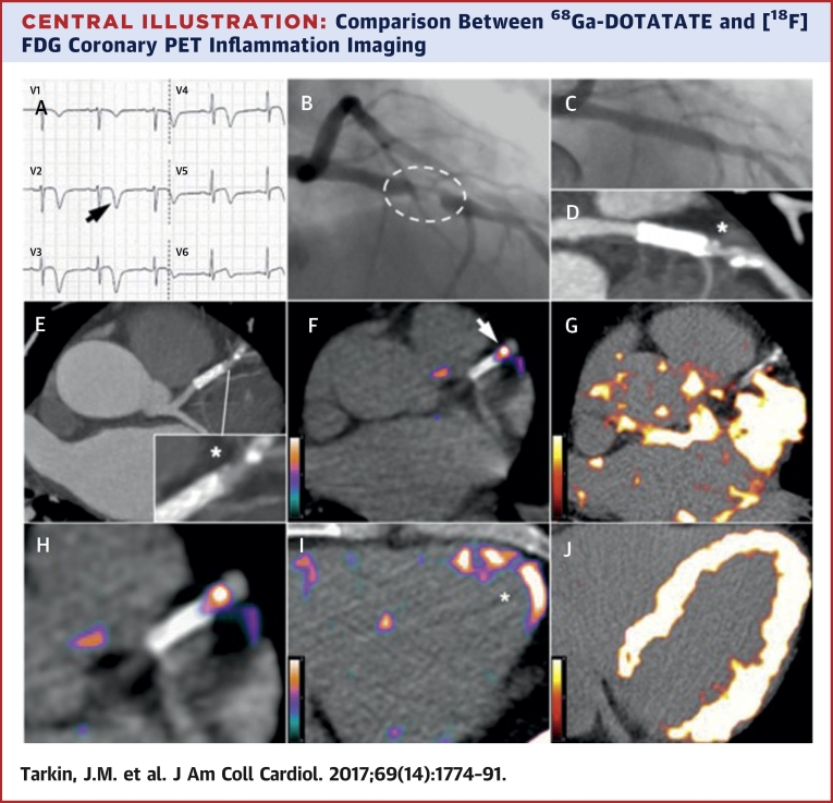
Inflammation drives atherosclerotic plaque rupture. Although inflammation can be measured using fluorine-18-labeled fluorodeoxyglucose positron emission tomography ([F]FDG PET), [F]FDG lacks cell specificity, and coronary imaging is unreliable because of myocardial spillover.
This study tested the efficacy of gallium-68-labeled DOTATATE (Ga-DOTATATE), a somatostatin receptor subtype-2 (SST)-binding PET tracer, for imaging atherosclerotic inflammation.
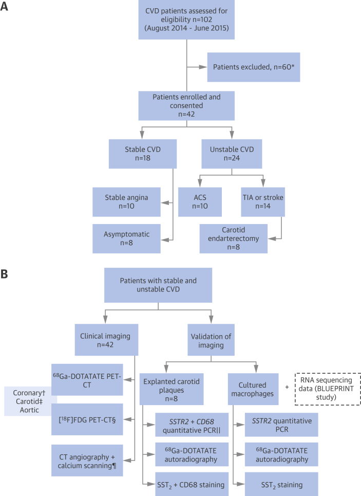
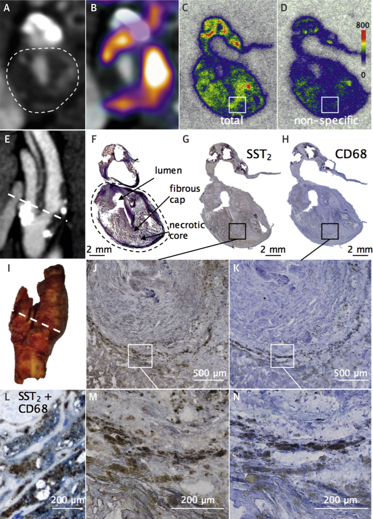
We confirmed Ga-DOTATATE binding in macrophages and excised carotid plaques. Ga-DOTATATE PET imaging was compared to [F]FDG PET imaging in 42 patients with atherosclerosis.
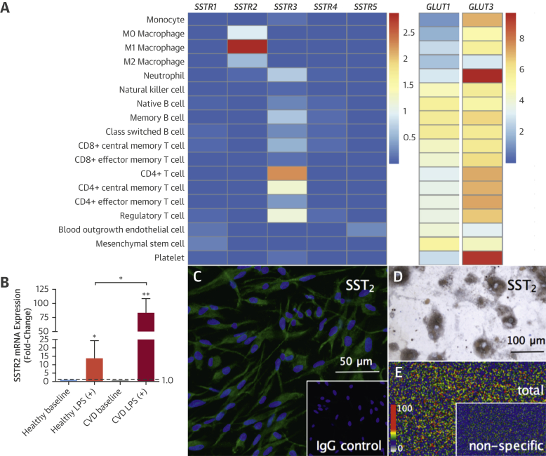
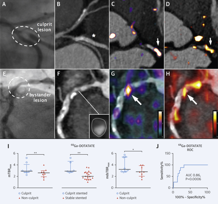
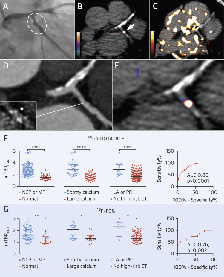
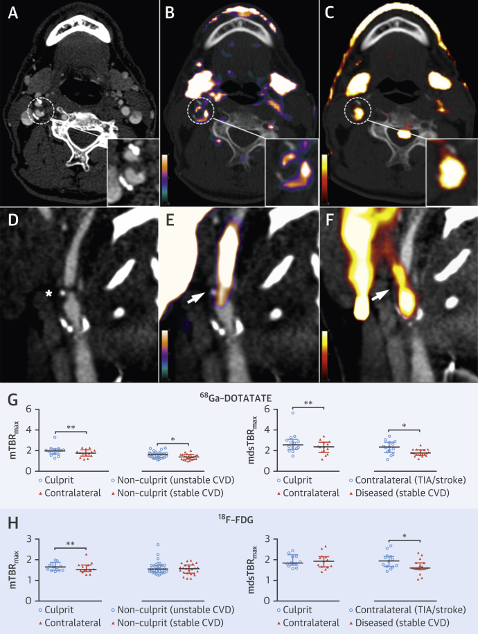
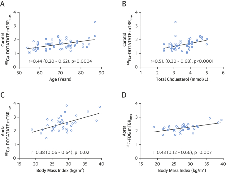
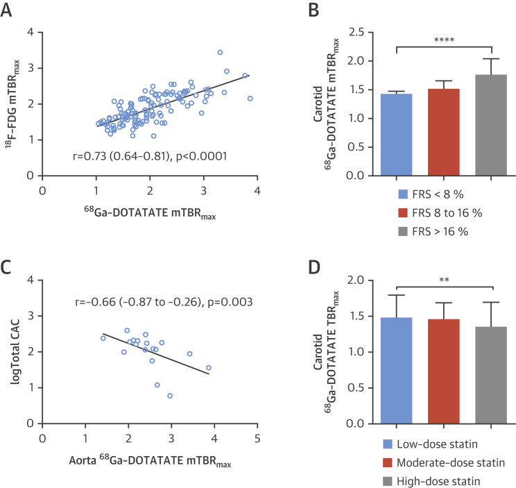
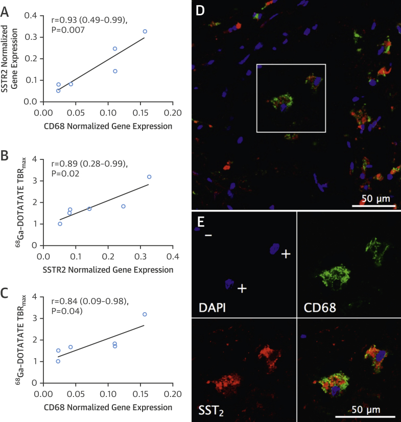
Target SSTR2 gene expression occurred exclusively in "proinflammatory" M1 macrophages, specific Ga-DOTATATE ligand binding to SST receptors occurred in CD68-positive macrophage-rich carotid plaque regions, and carotid SSTR2 mRNA was highly correlated with in vivo Ga-DOTATATE PET signals (r = 0.89; 95% confidence interval [CI]: 0.28 to 0.99; p = 0.02). Ga-DOTATATE mean of maximum tissue-to-blood ratios (mTBR) correctly identified culprit versus nonculprit arteries in patients with acute coronary syndrome (median difference: 0.69; interquartile range [IQR]: 0.22 to 1.15; p = 0.008) and transient ischemic attack/stroke (median difference: 0.13; IQR: 0.07 to 0.32; p = 0.003). Ga-DOTATATE mTBR predicted high-risk coronary computed tomography features (receiver operating characteristics area under the curve [ROC AUC]: 0.86; 95% CI: 0.80 to 0.92; p < 0.0001), and correlated with Framingham risk score (r = 0.53; 95% CI: 0.32 to 0.69; p <0.0001) and [F]FDG uptake (r = 0.73; 95% CI: 0.64 to 0.81; p < 0.0001). [F]FDG mTBR differentiated culprit from nonculprit carotid lesions (median difference: 0.12; IQR: 0.0 to 0.23; p = 0.008) and high-risk from lower-risk coronary arteries (ROC AUC: 0.76; 95% CI: 0.62 to 0.91; p = 0.002); however, myocardial [F]FDG spillover rendered coronary [F]FDG scans uninterpretable in 27 patients (64%). Coronary Ga-DOTATATE PET scans were readable in all patients.
We validated Ga-DOTATATE PET as a novel marker of atherosclerotic inflammation and confirmed that Ga-DOTATATE offers superior coronary imaging, excellent macrophage specificity, and better power to discriminate high-risk versus low-risk coronary lesions than [F]FDG. (Vascular Inflammation Imaging Using Somatostatin Receptor Positron Emission Tomography [VISION]; NCT02021188).
炎症促使动脉粥样硬化斑块破裂。虽然炎症可以通过氟-18标记的氟脱氧葡萄糖正电子发射断层扫描([F]FDG PET)来测量,但[F]FDG缺乏细胞特异性,并且由于心肌放射性外溢,冠状动脉成像不可靠。
本研究测试了镓-68标记的奥曲肽(Ga-DOTATATE),一种与生长抑素受体亚型2(SST)结合的PET示踪剂,用于成像动脉粥样硬化炎症的效果。
我们证实了Ga-DOTATATE在巨噬细胞和切除的颈动脉斑块中的结合。在42例动脉粥样硬化患者中,将Ga-DOTATATE PET成像与[F]FDG PET成像进行了比较。
目标SSTR2基因表达仅发生在“促炎”M1巨噬细胞中,特异性Ga-DOTATATE配体与SST受体的结合发生在富含CD68阳性巨噬细胞的颈动脉斑块区域,并且颈动脉SSTR2 mRNA与体内Ga-DOTATATE PET信号高度相关(r = 0.89;95%置信区间[CI]:0.28至0.99;p = 0.02)。Ga-DOTATATE的最大组织与血液比值平均值(mTBR)正确识别了急性冠状动脉综合征患者的罪犯血管与非罪犯血管(中位数差异:0.69;四分位间距[IQR]:0.22至1.15;p = 0.008)以及短暂性脑缺血发作/中风患者的罪犯血管与非罪犯血管(中位数差异:0.13;IQR:0.07至0.32;p = 0.003)。Ga-DOTATATE的mTBR预测了高危冠状动脉计算机断层扫描特征(受试者操作特征曲线下面积[ROC AUC]:0.86;95%CI:0.80至0.92;p < 0.0001),并且与弗雷明汉风险评分相关(r = 0.53;95%CI:0.32至0.69;p <0.0001)以及与[F]FDG摄取相关(r = 0.73;95%CI:0.64至0.81;p < 0.0001)。[F]FDG的mTBR区分了罪犯与非罪犯颈动脉病变(中位数差异:0.12;IQR:0.0至0.23;p = 0.008)以及高危与低危冠状动脉(ROC AUC:0.76;95%CI:0.62至0.91;p = 0.002);然而,心肌[F]FDG外溢使得27例患者(64%)的冠状动脉[F]FDG扫描无法解读。所有患者的冠状动脉Ga-DOTATATE PET扫描均可解读。
我们验证了Ga-DOTATATE PET作为动脉粥样硬化炎症的一种新型标志物,并证实Ga-DOTATATE在冠状动脉成像方面具有优势,具有出色的巨噬细胞特异性,并且与[F]FDG相比,在区分高危与低危冠状动脉病变方面具有更强的能力。(使用生长抑素受体正电子发射断层扫描的血管炎症成像[VISION];NCT02021188)