Khavandi Kaivan, Aghamohammadzadeh Reza, Luckie Matthew, Brownrigg Jack, Alam Uazman, Khattar Rajdeep, Malik Rayaz A, Heagerty Anthony M, Greenstein Adam S
Division of Cardiovascular Sciences, Manchester Academic Health Sciences Centre, University of Manchester, United Kingdom.
British Heart Foundation Centre of Excellence, The Rayne Institute, King's College London, London, United Kingdom.
J Am Heart Assoc. 2017 Apr 11;6(4):e004603. doi: 10.1161/JAHA.116.004603.
Small artery pathophysiology is frequently invoked as a cause of obesity-related diastolic heart failure. However, evidence to support this hypothesis is scant, particularly in humans.
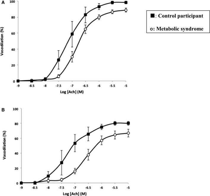
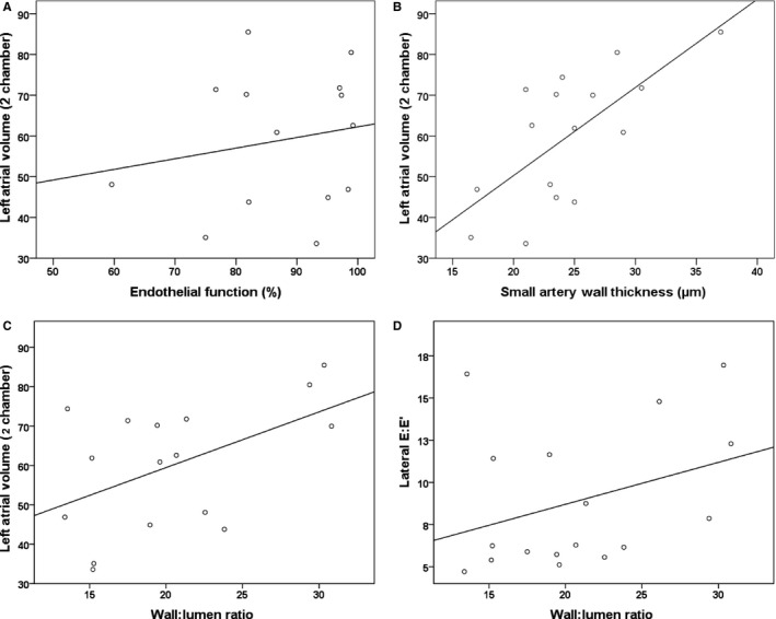
To address this, we studied human small artery structure and function in obesity and looked for correlations between vascular parameters and diastolic function. Seventeen obese patients with metabolic syndrome and 5 control participants underwent echocardiography and subcutaneous gluteal fat biopsy. Small arteries were isolated from the biopsy and pressure myography was used to study endothelial function and wall structure. In comparison with the control group, small arteries from obese participants exhibited significant endothelial dysfunction, assessed as the vasodilatory response to acetylcholine and also pathological growth of the wall. For the obese participants, multiple regression analysis revealed an association between left atrial volume and both the small artery wall thickness (β=0.718, =0.02) and wall-to-lumen ratio (β=0.605, =0.02). Furthermore, the E:E' ratio was associated with wall-to-lumen ratio (β=0.596, =0.02) and inversely associated with interleukin-6 (β=-0.868, =0.03). By contrast, endothelial function did not correlate with any of the echocardiographic parameters studied.
Although the small arteries studied were not cardiac in origin, our results support a role for small artery remodeling in the development of diastolic dysfunction in humans. Further direct examination of the structure and function of the myocardial resistance vasculature is now warranted, to elucidate the temporal association between metabolic risk factors, small artery injury, and diastolic impairment.
小动脉病理生理学常被认为是肥胖相关舒张性心力衰竭的一个病因。然而,支持这一假说的证据很少,尤其是在人类中。
为解决这一问题,我们研究了肥胖人群中小动脉的结构和功能,并寻找血管参数与舒张功能之间的相关性。17例患有代谢综合征的肥胖患者和5例对照参与者接受了超声心动图检查和臀下皮下脂肪活检。从活检组织中分离出小动脉,并用压力肌动描记法研究内皮功能和血管壁结构。与对照组相比,肥胖参与者的小动脉表现出明显的内皮功能障碍,通过对乙酰胆碱的血管舒张反应评估,同时血管壁出现病理性生长。对于肥胖参与者,多元回归分析显示左心房容积与小动脉壁厚度(β=0.718,P=0.02)和壁腔比(β=0.605,P=0.02)均相关。此外,E:E'比值与壁腔比相关(β=0.596,P=0.02),与白细胞介素-6呈负相关(β=-0.868,P=0.03)。相比之下,内皮功能与所研究的任何超声心动图参数均无相关性。
尽管所研究的小动脉并非源自心脏,但我们的结果支持小动脉重塑在人类舒张功能障碍发展中的作用。现在有必要进一步直接检查心肌阻力血管的结构和功能,以阐明代谢危险因素、小动脉损伤和舒张功能损害之间的时间关联。