Ruscitti Francesca, Ravanetti Francesca, Essers Jeroen, Ridwan Yanto, Belenkov Sasha, Vos Wim, Ferreira Francisca, KleinJan Alex, van Heijningen Paula, Van Holsbeke Cedric, Cacchioli Antonio, Villetti Gino, Stellari Franco Fabio
Chiesi S.p.A., Pre-Clinical R & D, Parma, Italy.
Dipartimento di Scienze Medico Veterinarie, Università di Parma, Parma, Italy.
Multidiscip Respir Med. 2017 Apr 10;12:8. doi: 10.1186/s40248-017-0089-0. eCollection 2017.
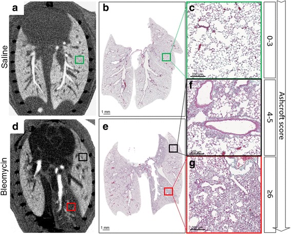
The intratracheal instillation of bleomycin in mice induces early damage to alveolar epithelial cells and development of inflammation followed by fibrotic tissue changes and represents the most widely used model of pulmonary fibrosis to investigate human IPF. Histopathology is the gold standard for assessing lung fibrosis in rodents, however it precludes repeated and longitudinal measurements of disease progression and does not provide information on spatial and temporal distribution of tissue damage. Here we investigated the use of the Micro-CT technique to allow the evaluation of disease onset and progression at different time-points in the mouse bleomycin model of lung fibrosis. Micro-CT was throughout coupled with histological analysis for the validation of the imaging results.
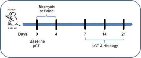
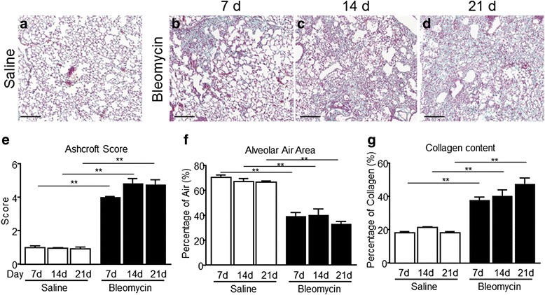
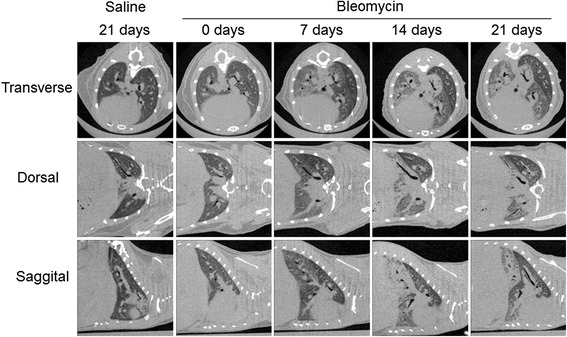
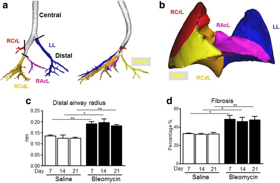
In bleomycin-instilled and control mice, airways and lung morphology changes were assessed and reconstructed at baseline, 7, 14 and 21 days post-treatment based on Micro-CT images. Ashcroft score, percentage of collagen content and percentage of alveolar air area were detected on lung slides processed by histology and subsequently compared with Micro-CT parameters.
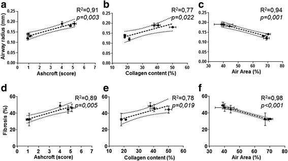
Extent (%) of fibrosis measured by Micro-CT correlated with Ashcroft score, the percentage of collagen content and the percentage of alveolar air area ( = 0.91; 0.77; 0.94, respectively). Distal airway radius also correlated with the Ashcroft score, the collagen content and alveolar air area percentage ( = 0.89; 0.78; 0.98, respectively).
Micro-CT data were in good agreement with histological read-outs as micro-CT was able to quantify effectively and non-invasively disease progression longitudinally and to reduce the variability and number of animals used to assess the damage. This suggests that this technique is a powerful tool for understanding experimental pulmonary fibrosis and that its use could translate into a more efficient drug discovery process, also helping to fill the gap between preclinical setting and clinical practice.
对小鼠气管内滴注博来霉素可导致肺泡上皮细胞早期损伤和炎症发展,随后出现纤维化组织改变,这是研究人类特发性肺纤维化(IPF)时最广泛使用的肺纤维化模型。组织病理学是评估啮齿动物肺纤维化的金标准,然而它无法对疾病进展进行重复和纵向测量,也不能提供有关组织损伤时空分布的信息。在此,我们研究了使用微型计算机断层扫描(Micro-CT)技术来评估肺纤维化小鼠模型在不同时间点的疾病发病和进展情况。整个实验过程中,Micro-CT均与组织学分析相结合以验证成像结果。
在接受博来霉素滴注的小鼠和对照小鼠中,基于Micro-CT图像在基线、治疗后7天、14天和21天评估并重建气道和肺形态变化。在经组织学处理的肺切片上检测阿什克罗夫特评分、胶原含量百分比和肺泡气腔面积百分比,随后与Micro-CT参数进行比较。
通过Micro-CT测量的纤维化程度(%)与阿什克罗夫特评分、胶原含量百分比和肺泡气腔面积百分比相关(分别为r = 0.91;0.77;0.94)。远端气道半径也与阿什克罗夫特评分、胶原含量和肺泡气腔面积百分比相关(分别为r = 0.89;0.78;0.98)。
Micro-CT数据与组织学读数高度一致,因为Micro-CT能够纵向有效地、非侵入性地量化疾病进展,并减少用于评估损伤的动物的变异性和数量。这表明该技术是理解实验性肺纤维化的有力工具,其应用可能转化为更高效的药物发现过程,也有助于填补临床前研究与临床实践之间的差距。