Cerny Milena, Omoumi Patrick, Larbi Ahmed, Manicourt Daniel, Perozziello Anne, Lecouvet Frederic E, Berg Bruno Vande, Dallaudière Benjamin
Department of Radiology, Cliniques Universitaires Saint-Luc, Université Catholique de Louvain, Brussel, Belgium.
Department of Radiology, University Hospital of Lausanne, Lausanne, Switzerland.
Eur J Radiol Open. 2017 Apr 2;4:40-44. doi: 10.1016/j.ejro.2017.03.002. eCollection 2017.
To determine if diagnostic signs of adhesive capsulitis (AC) of the shoulder at Magnetic Resonance Imaging (MRI) and arthrography (MRA) are applicable to CT arthrography (CTA).
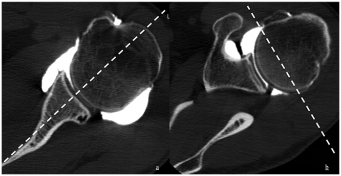
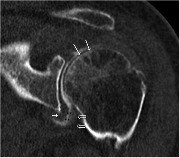
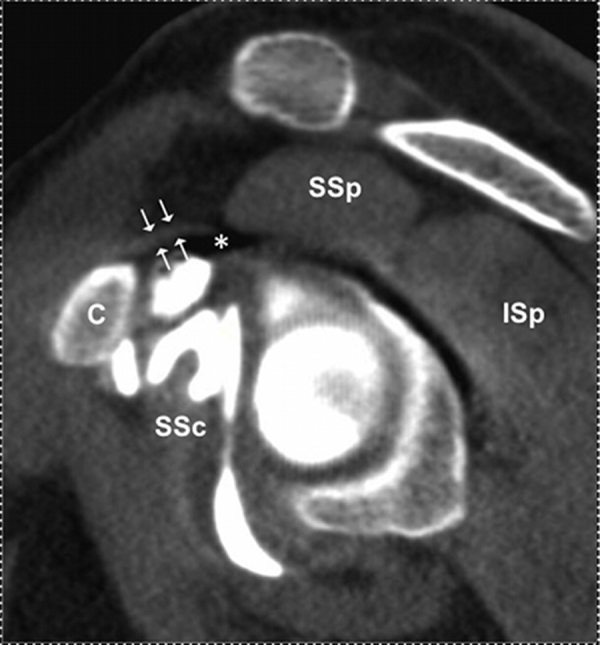
22 shoulder CTAs with AC were retrospectively reviewed for features described in MR literature. The control group was composed of 83 shoulder CTA divided into four subgroups 1) normal (N = 20), 2) omarthrosis (N = 19), 3) labral injury (N = 23), and 4) rotator cuff tear (N = 21). Two musculoskeletal radiologists assessed the rotator interval (RI) for obliteration, increased width and thickening of coracohumeral ligament (CHL). The width and capsule thickness of the axillary recess were measured.
The width of the axillary recess was significantly decreased in the AC group (4.6 ± 2.6 mm versus 9.9 ± 4.6 mm, p ≤ 0.0001; sensitivity and specificity of 84% and 80%). Thickness of the medial and lateral walls of the axillary capsule was significantly increased in the AC group (5.9 ± 1.3 mm versus 3.7 ± 1.1 mm, p ≤ 0.0001 and 5.7 ± 1 mm versus 3.5 ± 1.3 mm, p ≤ 0.0001, respectively). CHL thickness was significantly increased in the AC group (4.1 ± 1 mm (p ≤ 0.001)) in comparison to others groups. Obliteration of the RI was statistically significantly more frequent in patients with AC (72.7% (16/22) vs. 12% (10/83), p < 0.0001). Width of the RI did not differ significantly between patients and controls (p ≥ 0.428).
Decreased axillary width, and thickened axillary capsule are MR signs of AC applicable to CTA. Evaluation of rotator interval seems useful and reproducible only for obliteration.
确定磁共振成像(MRI)和关节造影(MRA)中肩关节粘连性关节囊炎(AC)的诊断征象是否适用于CT关节造影(CTA)。
对22例患有AC的肩部CTA进行回顾性分析,以查找MR文献中描述的特征。对照组由83例肩部CTA组成,分为四个亚组:1)正常组(N = 20),2)全关节病组(N = 19),3)盂唇损伤组(N = 23),4)肩袖撕裂组(N = 21)。两名肌肉骨骼放射科医生评估了旋转间隙(RI)是否闭塞、喙肱韧带(CHL)宽度增加和增厚情况。测量腋窝隐窝的宽度和关节囊厚度。
AC组腋窝隐窝宽度显著减小(4.6±2.6mm对9.9±4.6mm,p≤0.0001;敏感性和特异性分别为84%和80%)。AC组腋窝关节囊内侧壁和外侧壁厚度显著增加(分别为5.9±1.3mm对3.7±1.1mm,p≤0.0001;5.7±1mm对3.5±1.3mm,p≤0.0001)。与其他组相比,AC组CHL厚度显著增加(4.1±1mm(p≤0.001))。AC患者中RI闭塞在统计学上更为常见(72.7%(16/22)对12%(10/83),p<0.0001)。患者与对照组之间RI宽度差异无统计学意义(p≥0.428)。
腋窝宽度减小和腋窝关节囊增厚是适用于CTA的AC的MR征象。对旋转间隙的评估似乎仅对闭塞情况有用且可重复。