Kao Yu-Ting, Wu Chin-Han, Wu Shan-Ying, Lan Sheng-Hui, Liu Hsiao-Sheng, Tseng Ya-Shih
Department of Microbiology and Immunology, College of Medicine, National Cheng Kung University, Tainan, Taiwan.
Institute of Basic Medical Sciences, College of Medicine, National Cheng Kung University, Tainan, Taiwan.
BMC Cancer. 2017 Apr 18;17(1):277. doi: 10.1186/s12885-017-3253-1.
Arsenic is a widely distributed metalloid compound that has biphasic effects on cultured cells. In large doses, arsenic can be toxic enough to trigger cell death. In smaller amounts, non-toxic doses may promote cell proliferation and induces carcinogenesis. Aberration of chromosome is frequently detected in epithelial cells and lymphocytes of individuals from arsenic contaminated areas. Overexpression of Aurora-A, a mitotic kinase, results in chromosomal instability and cell transformation. We have reported that low concentration (≦1 μM) of arsenic treatment increases Aurora-A expression in immortalized bladder urothelial E7 cells. However, how arsenic induces carcinogenesis through Aurora-A activation remaining unclear.
Bromodeoxyuridine (BrdU) staining, MTT assay, and flow cytometry assay were conducted to determine cell proliferation. Messenger RNA and protein expression levels of Aurora-A were detected by reverse transcriptional-PCR and Western blotting, respectively. Centrosome of cells was observed by immunofluorescent staining. The transcription factor of Aurora-A was investigated by promoter activity, chromosome immunoprecipitation (ChIP), and small interfering RNA (shRNA) assays. Mouse model was utilized to confirm the relationship between arsenic and Aurora-A.
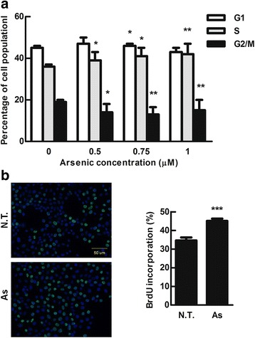
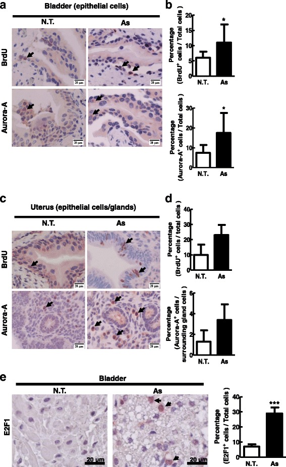
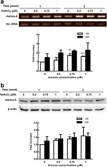
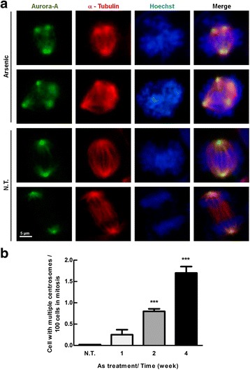
We reveal that low dosage of arsenic treatment increased cell proliferation is associated with accumulated cell population at S phase. We also detected increased Aurora-A expression at mRNA and protein levels in immortalized bladder urothelial E7 cells exposed to low doses of arsenic. Arsenic-treated cells displayed increased multiple centrosome which is resulted from overexpressed Aurora-A. Furthermore, the transcription factor, E2F1, is responsible for Aurora-A overexpression after arsenic treatment. We further disclosed that Aurora-A expression and cell proliferation were increased in bladder and uterus tissues of the BALB/c mice after long-term arsenic (1 mg/L) exposure for 2 months.
We reveal that low dose of arsenic induced cell proliferation is through Aurora-A overexpression, which is transcriptionally regulated by E2F1 both in vitro and in vivo. Our findings disclose a new possibility that arsenic at low concentration activates Aurora-A to induce carcinogenesis.
砷是一种广泛分布的类金属化合物,对培养细胞具有双相效应。大剂量时,砷毒性足以引发细胞死亡。小剂量时,无毒剂量可能促进细胞增殖并诱导致癌。在来自砷污染地区的个体的上皮细胞和淋巴细胞中经常检测到染色体畸变。有丝分裂激酶Aurora-A的过表达会导致染色体不稳定和细胞转化。我们曾报道,低浓度(≦1 μM)的砷处理会增加永生化膀胱尿路上皮E7细胞中Aurora-A的表达。然而,砷如何通过激活Aurora-A诱导致癌尚不清楚。
进行溴脱氧尿苷(BrdU)染色、MTT测定和流式细胞术测定以确定细胞增殖。分别通过逆转录PCR和蛋白质印迹检测Aurora-A的信使核糖核酸和蛋白质表达水平。通过免疫荧光染色观察细胞的中心体。通过启动子活性、染色体免疫沉淀(ChIP)和小干扰RNA(shRNA)测定研究Aurora-A的转录因子。利用小鼠模型证实砷与Aurora-A之间的关系。
我们发现低剂量砷处理增加细胞增殖与S期细胞群体积累有关。我们还检测到暴露于低剂量砷的永生化膀胱尿路上皮E7细胞中Aurora-A的信使核糖核酸和蛋白质水平表达增加。砷处理的细胞显示多个中心体增加,这是由Aurora-A过表达导致的。此外,转录因子E2F1负责砷处理后Aurora-A的过表达。我们进一步发现,长期暴露于砷(1 mg/L)2个月后BALB/c小鼠的膀胱和子宫组织中Aurora-A表达和细胞增殖增加。
我们发现低剂量砷诱导细胞增殖是通过Aurora-A过表达,其在体外和体内均受E2F1转录调控。我们的研究结果揭示了低浓度砷激活Aurora-A诱导致癌的新可能性。