Zhao Lei, Li Cheng, Liu Fei, Zhao Yonghong, Liu Jun, Hua Ye, Liu Jinyang, Huang Jiapeng, Ge Chunlin
Department of Pancreatic and Biliary Surgery, The First Hospital of China Medical University.
Department of Immunology, College of Basic Medical Sciences, China Medical University, Shenyang, Liaoning, People's Republic of China.
Onco Targets Ther. 2017 Apr 12;10:2115-2126. doi: 10.2147/OTT.S130481. eCollection 2017.
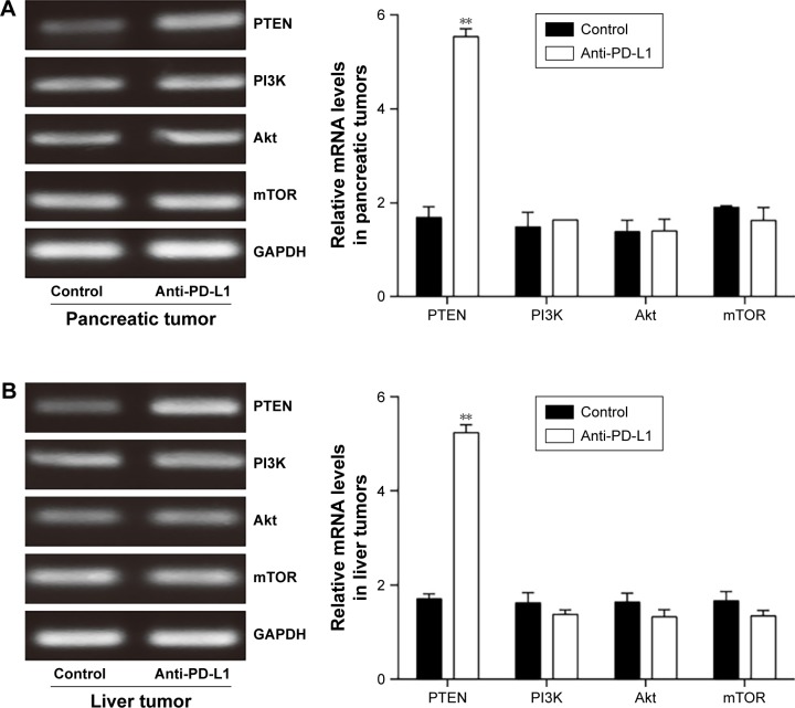
Pancreatic cancer is one of the most aggressive and intractable malignant tumors, and most deaths from pancreatic cancer are related to metastases. It has been demonstrated in vitro that overexpression of programmed death-ligand 1 (PD-L1) correlates with a lack of phosphatase and tensin homologue (PTEN) expression in pancreatic cancer tissue. This loss of PTEN expression may aberrantly activate the phosphatidylinositol 3-kinase (PI3K)/Akt/mammalian target of rapamycin (mTOR) pathway, and thereby promote tumor cell survival, proliferation, and disease progression. In this study, we investigated the potential therapeutic effect of blockading PD-L1 expression on the progression of pancreatic cancer and its spontaneous liver metastases in vivo by inhibiting the PI3K/Akt/mTOR signaling pathway.
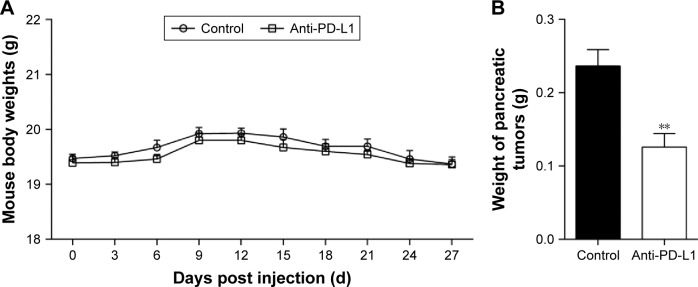
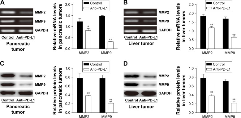
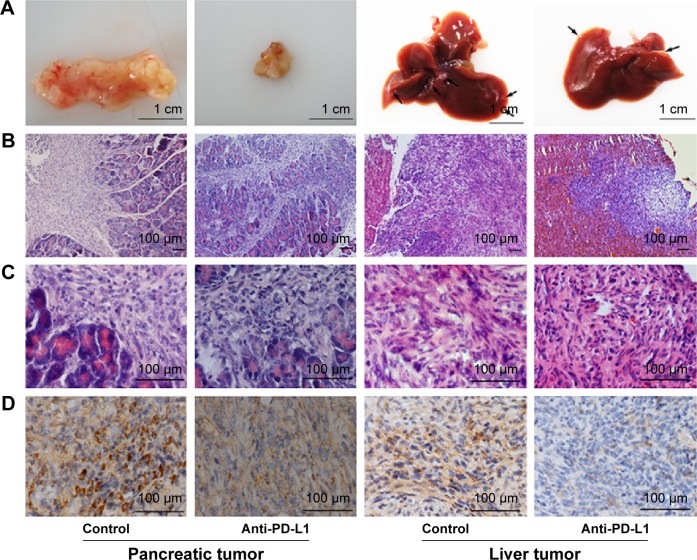
We investigated the effect of blockading PD-L1 in an orthotopic pancreatic cancer mouse model. The pancreatic tumor weights and inhibition ratios were determined after treatment with antimouse PD-L1 antibody for 5 weeks. We used immunohistochemistry methods to investigate PD-L1 expression in pancreatic cancer tissue and spontaneous liver metastasis tissue. The levels of mRNA and protein expression for various components involved in the PI3K/Akt/mTOR signaling pathway as well as for matrix metalloproteinases-2 and -9 (MMP2 and MMP9) were measured by reverse transcription polymerase chain reaction (RT-PCR) and Western blot methods, respectively.
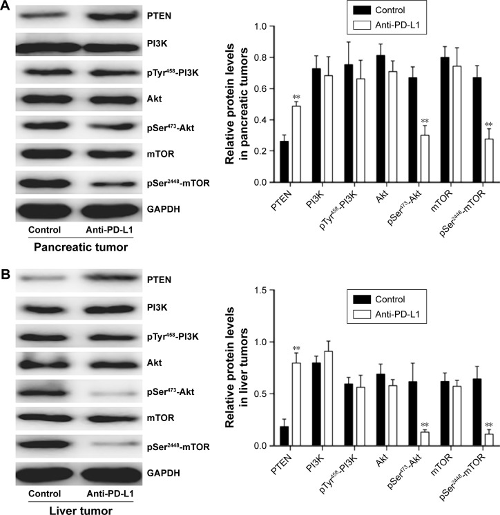
Blockading PD-L1 significantly inhibited tumor growth and decreased the levels of PD-L1 expression in tumor tissue. Furthermore, the levels of PTEN mRNA and protein expression were elevated, while the levels of phospho-Akt (p-Akt) and phospho-mTOR (p-mTOR) protein were decreased in pancreatic cancer and liver metastasis tissues after establishing a PD-L1 blockade. In addition, a PD-L1 blockade decreased the levels of MMP2 and MMP9 mRNA and protein expression in tumor tissues.
Our results suggest that a blockade of PD-L1 may inhibit the growth and metastasis of pancreatic cancer by modulating the PI3K/Akt/mTOR pathway.
胰腺癌是最具侵袭性和难治性的恶性肿瘤之一,大多数胰腺癌死亡与转移有关。体外研究表明,程序性死亡配体1(PD-L1)的过表达与胰腺癌组织中磷酸酶和张力蛋白同源物(PTEN)表达缺失相关。PTEN表达缺失可能异常激活磷脂酰肌醇3-激酶(PI3K)/蛋白激酶B(Akt)/雷帕霉素哺乳动物靶蛋白(mTOR)信号通路,从而促进肿瘤细胞存活、增殖及疾病进展。在本研究中,我们通过抑制PI3K/Akt/mTOR信号通路,研究阻断PD-L1表达对胰腺癌体内进展及其自发性肝转移的潜在治疗作用。
我们在原位胰腺癌小鼠模型中研究阻断PD-L1的作用。用抗小鼠PD-L1抗体治疗5周后,测定胰腺肿瘤重量和抑制率。我们采用免疫组织化学方法研究胰腺癌组织和自发性肝转移组织中PD-L1的表达。分别通过逆转录聚合酶链反应(RT-PCR)和蛋白质免疫印迹法检测PI3K/Akt/mTOR信号通路相关各组分以及基质金属蛋白酶-2和-9(MMP2和MMP9)的mRNA和蛋白表达水平。
阻断PD-L1可显著抑制肿瘤生长,并降低肿瘤组织中PD-L1表达水平。此外,在建立PD-L1阻断后,胰腺癌和肝转移组织中PTEN的mRNA和蛋白表达水平升高,而磷酸化Akt(p-Akt)和磷酸化mTOR(p-mTOR)蛋白水平降低。此外,阻断PD-L1可降低肿瘤组织中MMP2和MMP9的mRNA和蛋白表达水平。
我们的结果表明,阻断PD-L1可能通过调节PI3K/Akt/mTOR信号通路抑制胰腺癌的生长和转移。