Department of Plastic Surgery, BG University Hospital Bergmannsheil, Ruhr-University Bochum, Bochum, Germany.
Department of Plastic Surgery, BG Trauma Hospital Ludwigshafen, University of Heidelberg, Ludwigshafen, Germany.
J Cell Mol Med. 2017 Nov;21(11):2773-2781. doi: 10.1111/jcmm.13192. Epub 2017 Apr 26.
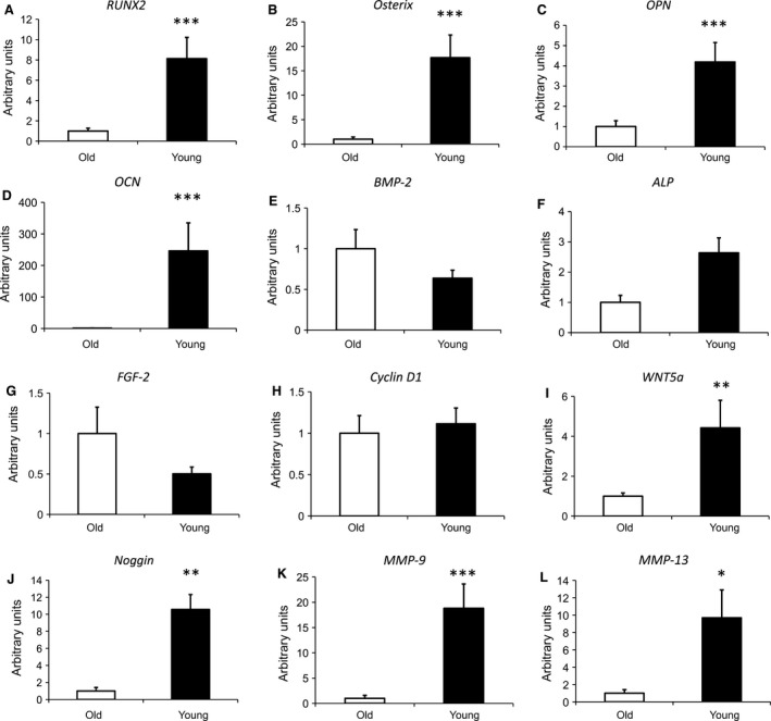
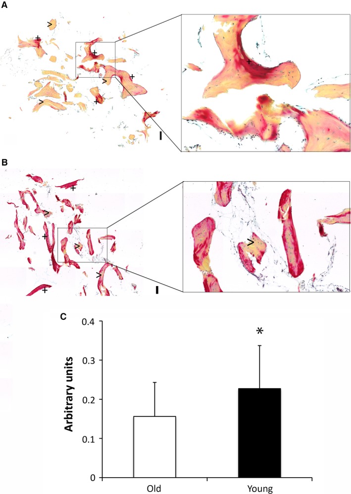
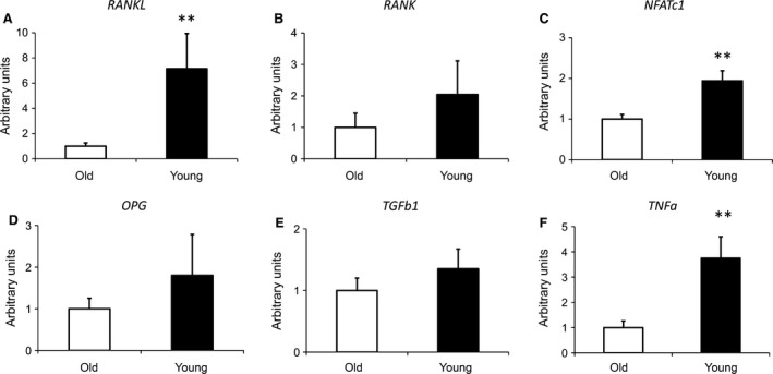
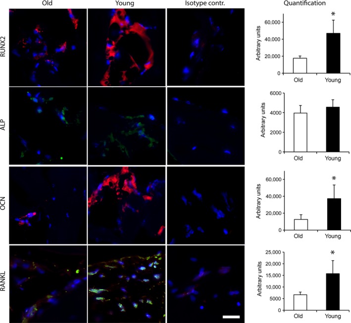
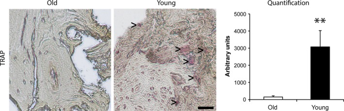
It is assumed that the activity of osteoblasts and osteoclasts is decreased in bone tissue of aged individuals. However, detailed investigation of the molecular signature of human bone from young compared to aged individuals confirming this assumption is lacking. In this study, quantitative expression analysis of genes related to osteogenesis and osteoclastogenesis of human cancellous bone derived from the distal radius of young and aged individuals was performed. Furthermore, we additionally performed immunohistochemical stainings. The young group included 24 individuals with an average age of 23.2 years, which was compared to cancellous bone derived from 11 body donators with an average age of 81.0 years. In cancellous bone of young individuals, the osteogenesis-related genes RUNX-2, OSTERIX, OSTEOPONTIN and OSTEOCALCIN were significantly up-regulated compared to aged individuals. In addition, RANKL and NFATc1, both markers for osteoclastogenesis, were significantly induced in cancellous bone of young individuals, as well as the WNT gene family member WNT5a and the matrix metalloproteinases MMP-9. However, quantitative RT-PCR analysis of BMP-2, ALP, FGF-2, CYCLIN-D1, MMP-13, RANK, OSTEOPROTEGERIN and TGFb1 revealed no significant difference. Furthermore, Tartrate-resistant acid phosphatase (TRAP) staining was performed which indicated an increased osteoclast activity in cancellous bone of young individuals. In addition, pentachrome stainings revealed significantly less mineralized bone matrix, more osteoid and an increased bone density in young individuals. In summary, markers related to osteogenesis as well as osteoclastogenesis were significantly decreased in the aged individuals. Thus, the present data extends the knowledge about reduced bone regeneration and healing capacity observed in aged individuals.
据假设,成骨细胞和破骨细胞的活性在老年人的骨组织中降低。然而,缺乏对来自年轻人和老年人的人松质骨的分子特征进行详细研究以证实这一假设。在这项研究中,对来自年轻人和老年人的人松质骨中成骨和破骨相关基因的定量表达分析进行了研究。此外,我们还进行了免疫组织化学染色。年轻人组包括 24 名平均年龄为 23.2 岁的个体,与平均年龄为 81.0 岁的 11 名遗体捐赠者的松质骨进行了比较。在年轻人的松质骨中,与老年人相比,成骨相关基因 RUNX-2、OSTERIX、骨桥蛋白和骨钙素明显上调。此外,RANKL 和 NFATc1,破骨细胞形成的两个标志物,在年轻人的松质骨中也明显被诱导,以及 WNT 基因家族成员 WNT5a 和基质金属蛋白酶 MMP-9。然而,BMP-2、ALP、FGF-2、CYCLIN-D1、MMP-13、RANK、骨保护素和 TGFb1 的定量 RT-PCR 分析显示没有明显差异。此外,进行了抗酒石酸酸性磷酸酶(TRAP)染色,表明年轻人的松质骨中破骨细胞活性增加。此外,五倍体染色显示年轻人的松质骨中矿化骨基质明显减少,类骨质增加,骨密度增加。总之,在老年人中,与成骨和破骨相关的标志物明显减少。因此,本研究扩展了关于老年人中观察到的骨再生和愈合能力降低的知识。