Blauwhoff-Buskermolen Susanne, Langius Jacqueline A E, Becker Annemarie, Verheul Henk M W, de van der Schueren Marian A E
Department of Nutrition and Dietetics, Internal Medicine, VU University Medical Center, Amsterdam, The Netherlands.
Department of Medical Oncology, VU University Medical Center, Amsterdam, The Netherlands.
J Cachexia Sarcopenia Muscle. 2017 Aug;8(4):615-622. doi: 10.1002/jcsm.12200. Epub 2017 Apr 26.
Progressive loss of muscle mass is a major characteristic of cancer cachexia. Consensus definitions for cachexia provide different options to measure muscle mass. This study describes the effect of different methods to determine muscle mass on the diagnosis of cancer cachexia. In addition, the association of cachexia with other features of cachexia, quality of life, and survival was explored.
Prior to chemotherapy, cachexia was assessed by weight loss, body mass index, and muscle mass measurements, the latter by mid-upper arm muscle area (MUAMA), computed tomography (CT) scans, and bio-electrical impedance analysis (BIA). In addition, appetite, inflammation, muscle strength, fatigue, quality of life, and survival were measured, and associations with cachexia were explored.
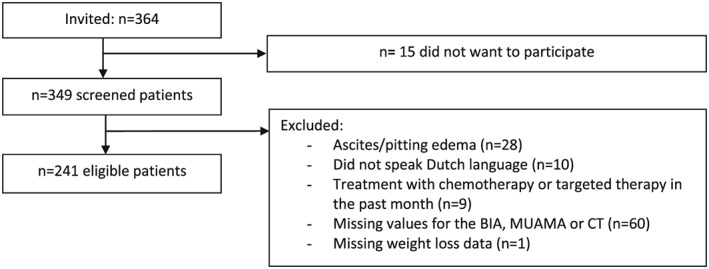
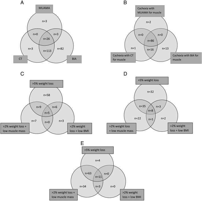
Included were 241 patients with advanced cancer of the lung (36%), colon/rectum (31%), prostate (18%), or breast (15%). Mean age was 64 ± 10 years; 54% was male. Prevalence of low muscle mass was as follows: 13% with MUAMA, 59% with CT, and 93% with BIA. In turn, the prevalence of cachexia was 37, 43, and 48%, whereby weight loss >5% was the most prominent component of being defined cachectic. Irrespective of type of muscle measurement, patients with cachexia presented more often with anorexia, inflammation, low muscle strength, and fatigue and had lower quality of life. Patients with cachexia had worse overall survival compared with patients without cachexia: HRs 2.00 (1.42-2.83) with MUAMA, 1.64 (1.15-2.34) with CT, and 1.50 (1.05-2.14) with BIA.
Although the prevalence of low muscle mass in patients with cancer depended largely on the type of muscle measurement, this had little influence on the diagnosis of cancer cachexia (as the majority of patients was already defined cachectic based on weight loss). New studies are warranted to further elucidate the additional role of muscle measurements in the diagnosis of cachexia and the association with clinical outcomes.
肌肉量的逐渐减少是癌症恶病质的主要特征。恶病质的共识定义提供了测量肌肉量的不同方法。本研究描述了不同肌肉量测定方法对癌症恶病质诊断的影响。此外,还探讨了恶病质与恶病质其他特征、生活质量及生存的相关性。
化疗前,通过体重减轻、体重指数和肌肉量测量评估恶病质,肌肉量测量采用上臂中部肌肉面积(MUAMA)、计算机断层扫描(CT)和生物电阻抗分析(BIA)。此外,还测量了食欲、炎症、肌肉力量、疲劳、生活质量和生存情况,并探讨了与恶病质的相关性。
纳入241例晚期癌症患者,其中肺癌患者占36%,结肠/直肠癌患者占31%,前列腺癌患者占18%,乳腺癌患者占15%。平均年龄为64±10岁;男性占54%。低肌肉量的患病率如下:MUAMA法为13%,CT法为59%,BIA法为93%。相应地,恶病质的患病率分别为37%、43%和48%,其中体重减轻>5%是定义为恶病质的最主要组成部分。无论采用何种肌肉测量方法,恶病质患者出现厌食、炎症、低肌肉力量和疲劳的情况更常见,生活质量更低。与无恶病质患者相比,恶病质患者的总生存期更差:MUAMA法的风险比为2.00(1.42 - 2.83),CT法为1.64(1.15 - 2.34),BIA法为1.50(1.05 - 2.14)。
虽然癌症患者低肌肉量的患病率在很大程度上取决于肌肉测量方法,但这对癌症恶病质的诊断影响不大(因为大多数患者已根据体重减轻被定义为恶病质)。有必要开展新的研究,以进一步阐明肌肉测量在恶病质诊断中的额外作用以及与临床结局的相关性。