Hirano Hirofumi, Kawahara Takashi, Niiro Masaki, Yonezawa Hajime, Takajyou Tomoko, Ohi Yasuyo, Kitazono Ikumi, Sakae Kiyohiro, Arita Kazunori
Department of Neurosurgery, Kagoshima University Graduate School of Medical and Dental Sciences, Kagoshima 890-8520, Japan.
Department of Neurosurgery, Imamura Bun-in Hospital, Kagoshima 890-0064, Japan.
Mol Clin Oncol. 2017 Mar;6(3):321-326. doi: 10.3892/mco.2017.1160. Epub 2017 Feb 7.
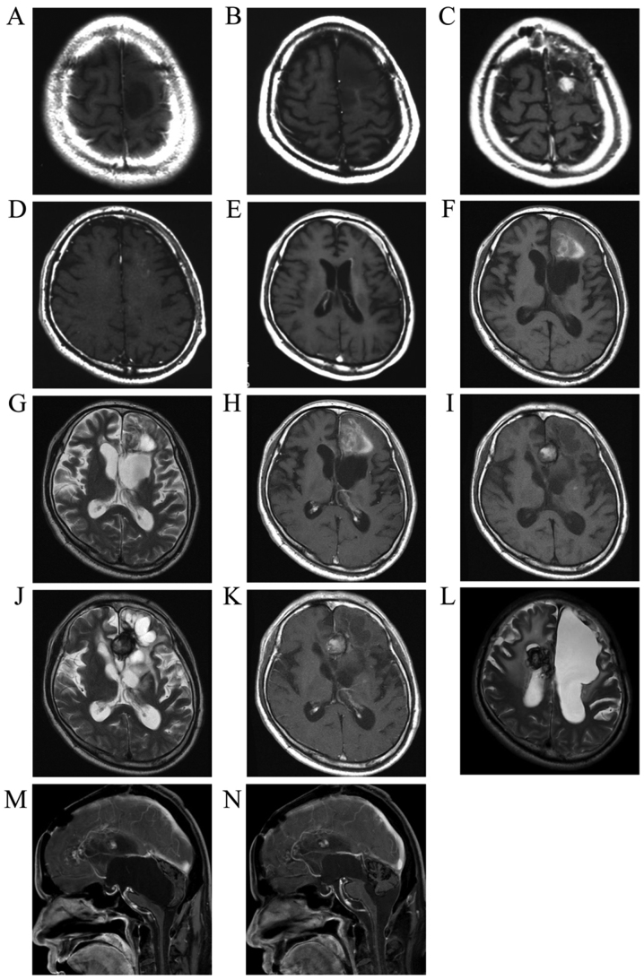
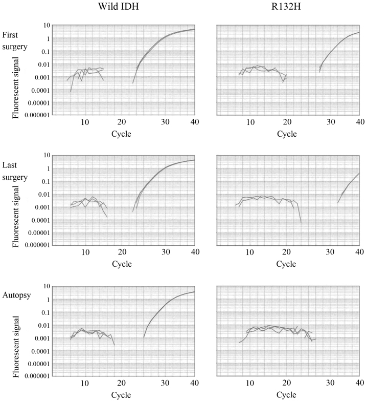
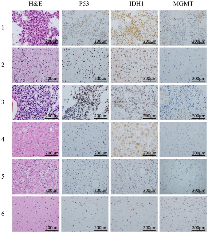
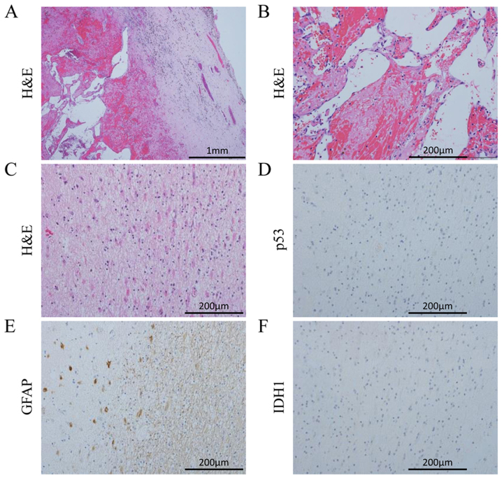
We herein present an autopsy case of a glioma patient who received long-term treatment with temozolomide (TMZ). The patient, a 35-year-old man with a hypointense tumor of the left frontal lobe, without contrast enhancement following gadolinium (Gd) administration on T1-weighted images, underwent tumor removal surgery, after which the tumor was diagnosed as anaplastic astrocytoma. By the third round of surgery, the tumor had progressed to anaplastic astrocytoma with contrast enhancement following Gd administration, and the patient received 60 Gy of external beam radiotherapy and nimustine hydrochloride (ACNU)-based chemotherapy. After the fifth tumor removal surgery, TMZ was substituted with ACNU chemotherapy, which suppressed tumor progression. Following the 41st TMZ treatment, hemorrhage was observed in the residual tumor, and the hematoma had been replaced by a hemangioma. The hemangioma and surrounding brain tissue was removed during the sixth surgery. The patient survived for 14 years and 9 months after the initial surgery, but succumbed to hydrocephalus due to bleeding from hemangiomas. The histopathological specimens of the first to the sixth surgeries revealed mutant isocitrate dehydrogenase 1 (IDH1; R132H point mutation) and p53-positive tumor cells, but cells positive for the R132H mutation or p53 could not be detected by immunohistochemistry in the autopsy specimens of the brain after 108 courses of TMZ treatment. Mutant IDH1 (R132H) cells were also not detected in the autopsy specimens of the brain by polymerase chain reaction analysis.
我们在此报告一例接受替莫唑胺(TMZ)长期治疗的胶质瘤患者的尸检病例。该患者为一名35岁男性,左额叶有低信号肿瘤,在T1加权图像上静脉注射钆(Gd)后无强化,接受了肿瘤切除术,术后肿瘤被诊断为间变性星形细胞瘤。到第三次手术时,肿瘤已进展为Gd注射后有强化的间变性星形细胞瘤,患者接受了60 Gy的外照射放疗和基于盐酸尼莫司汀(ACNU)的化疗。在第五次肿瘤切除术后,TMZ被ACNU化疗替代,这抑制了肿瘤进展。在第41次TMZ治疗后,在残留肿瘤中观察到出血,血肿已被血管瘤取代。在第六次手术中切除了血管瘤和周围脑组织。患者在初次手术后存活了14年9个月,但因血管瘤出血导致脑积水而死亡。第一次至第六次手术的组织病理学标本显示存在异柠檬酸脱氢酶1(IDH1;R132H点突变)突变和p53阳性肿瘤细胞,但在接受108个疗程TMZ治疗后的脑尸检标本中,通过免疫组织化学未检测到R132H突变或p53阳性的细胞。通过聚合酶链反应分析,在脑尸检标本中也未检测到突变的IDH1(R132H)细胞。