• 文献检索
  • 文档翻译
  • 深度研究
  • 学术资讯
  • Suppr Zotero 插件Zotero 插件
  • 邀请有礼
  • 套餐&价格
  • 历史记录
应用&插件
Suppr Zotero 插件Zotero 插件浏览器插件Mac 客户端Windows 客户端微信小程序
定价
高级版会员购买积分包购买API积分包
服务
文献检索文档翻译深度研究API 文档MCP 服务
关于我们
关于 Suppr公司介绍联系我们用户协议隐私条款
关注我们

Suppr 超能文献

核心技术专利:CN118964589B侵权必究
粤ICP备2023148730 号-1Suppr @ 2026

文献检索

告别复杂PubMed语法,用中文像聊天一样搜索,搜遍4000万医学文献。AI智能推荐,让科研检索更轻松。

立即免费搜索

文件翻译

保留排版,准确专业,支持PDF/Word/PPT等文件格式,支持 12+语言互译。

免费翻译文档

深度研究

AI帮你快速写综述,25分钟生成高质量综述,智能提取关键信息,辅助科研写作。

立即免费体验

髓核外植体中脊索细胞的再生潜能

The Regenerative Potential of Notochordal Cells in a Nucleus Pulposus Explant.

作者信息

Arkesteijn Irene T M, Potier Esther, Ito Keita

机构信息

Department of Biomedical Engineering, Eindhoven University of Technology, Eindhoven, The Netherlands.

Department of Osteoarticular Bioengineering and Bioimaging, University Paris Diderot, Paris, France.

出版信息

Global Spine J. 2017 Feb;7(1):14-20. doi: 10.1055/s-0036-1583174. Epub 2017 Feb 1.

DOI:10.1055/s-0036-1583174
PMID:28451504
原文链接:https://pmc.ncbi.nlm.nih.gov/articles/PMC5400162/
Abstract

STUDY DESIGN

In vitro disk explant culture.

OBJECTIVE

Notochordal cells (NCs) have been shown to upregulate matrix production by nucleus pulposus (NP) cells in coculture. To examine the translation of these in vitro results to a nativelike setting, the regenerative potential of NCs injected into NP tissue was assessed in this study.

METHODS

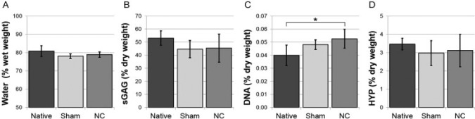
NP explants were cultured after injection with NCs in phosphate-buffered saline (PBS) or with PBS alone (sham). At days 0 and 42, cell viability and morphology, water, DNA, sulfated glycosaminoglycan and hydroxyproline content, and gene expression of anabolic markers were analyzed.

RESULTS

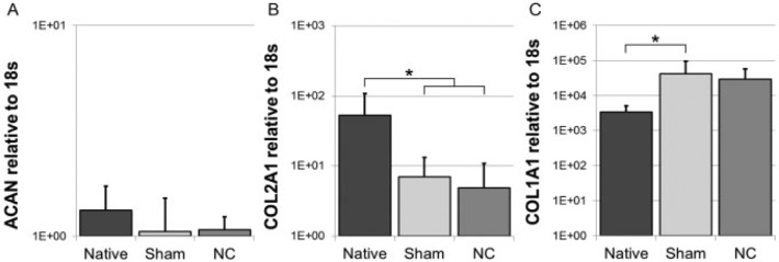
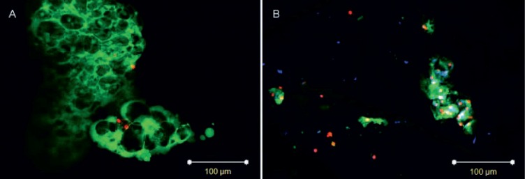
NCs remained viable during culture, but their morphology changed. The biochemical content remained unchanged, except for the DNA content in the NC group. Overall expression remained unchanged, whereas decreased during culture.

CONCLUSIONS

No overall anabolic response was observed when NCs were injected into NP explants. NCs were found to survive but did not display the typical NC morphology by the end of the culture period.

摘要

研究设计

体外椎间盘外植体培养。

目的

在共培养中,脊索细胞(NCs)已被证明可上调髓核(NP)细胞的基质产生。为了检验这些体外实验结果在类似天然环境中的转化情况,本研究评估了注入NP组织的NCs的再生潜力。

方法

NP外植体在注射含NCs的磷酸盐缓冲盐水(PBS)或仅注射PBS(假手术组)后进行培养。在第0天和第42天,分析细胞活力和形态、水、DNA、硫酸化糖胺聚糖和羟脯氨酸含量以及合成代谢标志物的基因表达。

结果

NCs在培养过程中保持存活,但其形态发生了变化。除NC组的DNA含量外,生化成分保持不变。总体表达保持不变,而在培养过程中下降。

结论

将NCs注入NP外植体时未观察到总体合成代谢反应。发现NCs存活,但在培养期结束时未表现出典型的NC形态。

https://cdn.ncbi.nlm.nih.gov/pmc/blobs/7420/5400162/1d26b583ef84/10.1055_s-0036-1583174-fig4.jpg
https://cdn.ncbi.nlm.nih.gov/pmc/blobs/7420/5400162/c38cee83eb41/10.1055_s-0036-1583174-fig1.jpg
https://cdn.ncbi.nlm.nih.gov/pmc/blobs/7420/5400162/1531a33af6d4/10.1055_s-0036-1583174-fig2.jpg
https://cdn.ncbi.nlm.nih.gov/pmc/blobs/7420/5400162/11400a26bd54/10.1055_s-0036-1583174-fig3.jpg
https://cdn.ncbi.nlm.nih.gov/pmc/blobs/7420/5400162/1d26b583ef84/10.1055_s-0036-1583174-fig4.jpg

相似文献

1
The Regenerative Potential of Notochordal Cells in a Nucleus Pulposus Explant.髓核外植体中脊索细胞的再生潜能
Global Spine J. 2017 Feb;7(1):14-20. doi: 10.1055/s-0036-1583174. Epub 2017 Feb 1.
2
Effect of coculturing canine notochordal, nucleus pulposus and mesenchymal stromal cells for intervertebral disc regeneration.共培养犬脊索细胞、髓核细胞和间充质基质细胞用于椎间盘再生的效果
Arthritis Res Ther. 2015 Mar 14;17(1):60. doi: 10.1186/s13075-015-0569-6.
3
Co-culturing nucleus pulposus mesenchymal stem cells with notochordal cell-rich nucleus pulposus explants attenuates tumor necrosis factor-α-induced senescence.将纤维环间充质干细胞与富含脊索细胞的纤维环髓核组织块共培养可减轻肿瘤坏死因子-α诱导的衰老。
Stem Cell Res Ther. 2018 Jun 26;9(1):171. doi: 10.1186/s13287-018-0919-9.
4
Coculture with Partial Digestion Notochordal Cell-Rich Nucleus Pulposus Tissue Activates Degenerative Human Nucleus Pulposus Cells.与部分消化的富含脊索细胞的髓核组织共培养可激活退变的人髓核细胞。
Tissue Eng Part A. 2017 Aug;23(15-16):837-846. doi: 10.1089/ten.TEA.2016.0428. Epub 2017 Feb 28.
5
The effect of a cyclooxygenase 2 inhibitor on early degenerated human nucleus pulposus explants.环氧化酶 2 抑制剂对早期退变人髓核组织块的作用。
Global Spine J. 2014 Feb;4(1):33-40. doi: 10.1055/s-0033-1359724. Epub 2013 Oct 23.
6
Native nucleus pulposus tissue matrix promotes notochordal differentiation of human induced pluripotent stem cells with potential for treating intervertebral disc degeneration.天然髓核组织基质可促进人诱导多能干细胞的脊索分化,具有治疗椎间盘退变的潜力。
J Biomed Mater Res A. 2015 Mar;103(3):1053-9. doi: 10.1002/jbm.a.35243. Epub 2014 Jun 17.
7
Increased osmolarity and cell clustering preserve canine notochordal cell phenotype in culture.渗透压升高和细胞聚集可在培养中保留犬脊索细胞表型。
Tissue Eng Part C Methods. 2014 Aug;20(8):652-62. doi: 10.1089/ten.TEC.2013.0479. Epub 2014 Feb 14.
8
The Stimulatory Effect of Notochordal Cell-Conditioned Medium in a Nucleus Pulposus Explant Culture.脊索细胞条件培养液在髓核组织块培养中的刺激作用。
Tissue Eng Part A. 2016 Jan;22(1-2):103-10. doi: 10.1089/ten.TEA.2015.0121. Epub 2015 Nov 5.
9
Osteogenic protein 1 does not stimulate a regenerative effect in cultured human degenerated nucleus pulposus tissue.成骨蛋白1对培养的人退变髓核组织无再生刺激作用。
J Tissue Eng Regen Med. 2017 Jul;11(7):2127-2135. doi: 10.1002/term.2111. Epub 2015 Nov 27.
10
Notochordal Cell Matrix As a Therapeutic Agent for Intervertebral Disc Regeneration.脊索细胞基质作为一种治疗椎间盘再生的治疗剂。
Tissue Eng Part A. 2019 Jun;25(11-12):830-841. doi: 10.1089/ten.TEA.2018.0026. Epub 2018 Jun 29.

引用本文的文献

1
Recommendations for intervertebral disc notochordal cell investigation: From isolation to characterization.椎间盘脊索细胞研究建议:从分离到表征
JOR Spine. 2023 Jul 9;6(3):e1272. doi: 10.1002/jsp2.1272. eCollection 2023 Sep.
2
Cell sources proposed for nucleus pulposus regeneration.被提议用于髓核再生的细胞来源。
JOR Spine. 2021 Nov 24;4(4):e1175. doi: 10.1002/jsp2.1175. eCollection 2021 Dec.
3
The Cellular Composition of Bovine Coccygeal Intervertebral Discs: A Comprehensive Single-Cell RNAseq Analysis.牛尾骨椎间盘中的细胞组成:全面的单细胞 RNAseq 分析。

本文引用的文献

1
A Well-Controlled Nucleus Pulposus Tissue Culture System with Injection Port for Evaluating Regenerative Therapies.一种带有注射端口的用于评估再生疗法的良好控制的髓核组织培养系统。
Ann Biomed Eng. 2016 May;44(5):1798-807. doi: 10.1007/s10439-015-1428-y. Epub 2015 Aug 21.
2
Conditioned medium derived from notochordal cell-rich nucleus pulposus tissue stimulates matrix production by canine nucleus pulposus cells and bone marrow-derived stromal cells.源自富含脊索细胞的髓核组织的条件培养基可刺激犬髓核细胞和骨髓源性基质细胞产生基质。
Tissue Eng Part A. 2015 Mar;21(5-6):1077-84. doi: 10.1089/ten.TEA.2014.0309. Epub 2014 Dec 17.
3
Int J Mol Sci. 2021 May 6;22(9):4917. doi: 10.3390/ijms22094917.
4
Multiscale Regulation of the Intervertebral Disc: Achievements in Experimental, In Silico, and Regenerative Research.椎间盘的多尺度调节:实验、计算机模拟和再生研究的成果
Int J Mol Sci. 2021 Jan 12;22(2):703. doi: 10.3390/ijms22020703.
5
LncRNA MALAT1 exhibits positive effects on nucleus pulposus cell biology in vivo and in vitro by sponging miR-503.长链非编码 RNA MALAT1 通过海绵吸附 miR-503 在体内和体外对髓核细胞生物学表现出正向作用。
BMC Mol Cell Biol. 2020 Mar 30;21(1):23. doi: 10.1186/s12860-020-00265-2.
6
Leaping the hurdles in developing regenerative treatments for the intervertebral disc from preclinical to clinical.跨越椎间盘再生治疗从临床前到临床阶段的障碍。
JOR Spine. 2018 Aug 2;1(3):e1027. doi: 10.1002/jsp2.1027. eCollection 2018 Sep.
7
Intervertebral Disc Nucleus Repair: Hype or Hope?椎间盘核修复:炒作还是希望?
Int J Mol Sci. 2019 Jul 24;20(15):3622. doi: 10.3390/ijms20153622.
8
Current strategies for treatment of intervertebral disc degeneration: substitution and regeneration possibilities.当前椎间盘退变的治疗策略:替代与再生的可能性
Biomater Res. 2017 Oct 23;21:22. doi: 10.1186/s40824-017-0106-6. eCollection 2017.
Potential application of notochordal cells for intervertebral disc regeneration: an in vitro assessment.
脊索细胞在椎间盘再生中的潜在应用:一项体外评估
Eur Cell Mater. 2014 Jul 25;28:68-80; discussion 80-1. doi: 10.22203/ecm.v028a06.
4
Increased osmolarity and cell clustering preserve canine notochordal cell phenotype in culture.渗透压升高和细胞聚集可在培养中保留犬脊索细胞表型。
Tissue Eng Part C Methods. 2014 Aug;20(8):652-62. doi: 10.1089/ten.TEC.2013.0479. Epub 2014 Feb 14.
5
Variations in gene and protein expression in canine chondrodystrophic nucleus pulposus cells following long-term three-dimensional culture.犬软骨发育不良髓核细胞长期三维培养后基因和蛋白质表达的变化。
PLoS One. 2013 May 2;8(5):e63120. doi: 10.1371/journal.pone.0063120. Print 2013.
6
Dose- and time-dependent effect of high glucose concentration on viability of notochordal cells and expression of matrix degrading and fibrotic enzymes.高糖浓度对脊索细胞活力及基质降解和纤维化酶表达的剂量和时间依赖性影响。
Int Orthop. 2013 Jun;37(6):1179-86. doi: 10.1007/s00264-013-1836-2. Epub 2013 Mar 16.
7
Long-term culture of bovine nucleus pulposus explants in a native environment.牛髓核组织在天然环境中的长期培养。
Spine J. 2013 Apr;13(4):454-63. doi: 10.1016/j.spinee.2012.12.006. Epub 2013 Jan 20.
8
The evolutionary importance of cell ratio between notochordal and nucleus pulposus cells: an experimental 3-D co-culture study.脊索细胞与髓核细胞比例的进化重要性:一项实验性三维共培养研究。
Eur Spine J. 2012 Aug;21 Suppl 6(Suppl 6):S819-25. doi: 10.1007/s00586-011-2026-9. Epub 2011 Sep 28.
9
Regenerative potential of TGFβ3 + Dex and notochordal cell conditioned media on degenerated human intervertebral disc cells.TGFβ3 + Dex 和脊索细胞条件培养基对退变人椎间盘细胞的再生潜能。
J Orthop Res. 2012 Mar;30(3):482-8. doi: 10.1002/jor.21534. Epub 2011 Aug 22.
10
Culturing bovine nucleus pulposus explants by balancing medium osmolarity.通过平衡培养基渗透压培养牛髓核组织块。
Tissue Eng Part C Methods. 2011 Nov;17(11):1089-96. doi: 10.1089/ten.TEC.2011.0215. Epub 2011 Aug 29.