• 文献检索
  • 文档翻译
  • 深度研究
  • 学术资讯
  • Suppr Zotero 插件Zotero 插件
  • 邀请有礼
  • 套餐&价格
  • 历史记录
应用&插件
Suppr Zotero 插件Zotero 插件浏览器插件Mac 客户端Windows 客户端微信小程序
定价
高级版会员购买积分包购买API积分包
服务
文献检索文档翻译深度研究API 文档MCP 服务
关于我们
关于 Suppr公司介绍联系我们用户协议隐私条款
关注我们

Suppr 超能文献

核心技术专利:CN118964589B侵权必究
粤ICP备2023148730 号-1Suppr @ 2026

文献检索

告别复杂PubMed语法,用中文像聊天一样搜索,搜遍4000万医学文献。AI智能推荐,让科研检索更轻松。

立即免费搜索

文件翻译

保留排版,准确专业,支持PDF/Word/PPT等文件格式,支持 12+语言互译。

免费翻译文档

深度研究

AI帮你快速写综述,25分钟生成高质量综述,智能提取关键信息,辅助科研写作。

立即免费体验

通过超分辨率显微镜剖析活细胞中的肌动蛋白皮质密度和膜-皮质距离。

Dissecting the actin cortex density and membrane-cortex distance in living cells by super-resolution microscopy.

作者信息

Clausen M P, Colin-York H, Schneider F, Eggeling C, Fritzsche M

机构信息

MRC Human Immunology Unit, Weatherall Institute of Molecular Medicine, University of Oxford, Headley Way, OX3 9DS Oxford, UK.

Department of Physics, Chemistry, and Pharmacy, MEMPHYS-Center for Biomembrane Physics, University of Southern Denmark, Campusvej 55, 5230 Odense M, Denmark.

出版信息

J Phys D Appl Phys. 2017 Feb 15;50(6):064002. doi: 10.1088/1361-6463/aa52a1. Epub 2017 Jan 11.

DOI:10.1088/1361-6463/aa52a1
PMID:28458398
原文链接:https://pmc.ncbi.nlm.nih.gov/articles/PMC5390943/
Abstract

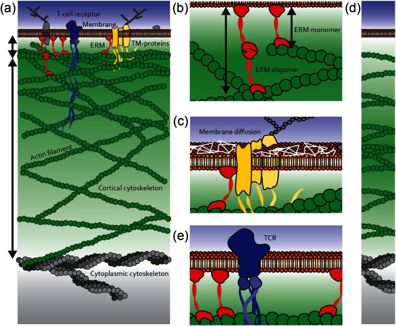
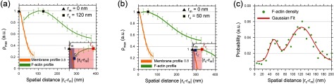
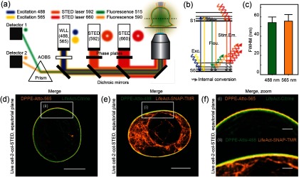
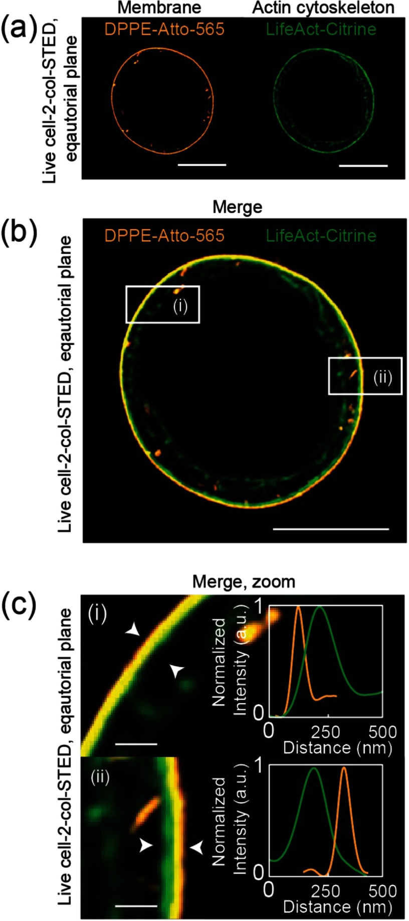
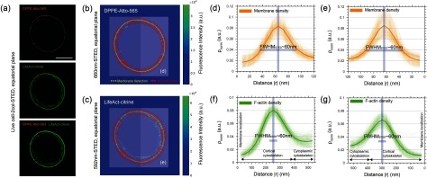
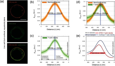
Nanoscale spacing between the plasma membrane and the underlying cortical actin cytoskeleton profoundly modulates cellular morphology, mechanics, and function. Measuring this distance has been a key challenge in cell biology. Current methods for dissecting the nanoscale spacing either limit themselves to complex survey design using fixed samples or rely on diffraction-limited fluorescence imaging whose spatial resolution is insufficient to quantify distances on the nanoscale. Using dual-color super-resolution STED (stimulated-emission-depletion) microscopy, we here overcome this challenge and accurately measure the density distribution of the cortical actin cytoskeleton and the distance between the actin cortex and the membrane in live Jurkat T-cells. We found an asymmetric cortical actin density distribution with a mean width of 230 (+105/-125) nm. The spatial distances measured between the maximum density peaks of the cortex and the membrane were bi-modally distributed with mean values of 50  ±  15 nm and 120  ±  40 nm, respectively. Taken together with the finite width of the cortex, our results suggest that in some regions the cortical actin is closer than 10 nm to the membrane and a maximum of 20 nm in others.

摘要

质膜与下方的皮质肌动蛋白细胞骨架之间的纳米级间距深刻地调节着细胞形态、力学和功能。测量这一距离一直是细胞生物学中的一项关键挑战。目前用于剖析纳米级间距的方法要么局限于使用固定样本的复杂测量设计,要么依赖于衍射极限荧光成像,其空间分辨率不足以量化纳米级的距离。在这里,我们使用双色超分辨率受激发射损耗(STED)显微镜克服了这一挑战,并准确测量了活的Jurkat T细胞中皮质肌动蛋白细胞骨架的密度分布以及肌动蛋白皮质与细胞膜之间的距离。我们发现皮质肌动蛋白密度分布不对称,平均宽度为230(+105/-125)nm。皮质与细胞膜的最大密度峰之间测得的空间距离呈双峰分布,平均值分别为50 ± 15 nm和120 ± 40 nm。结合皮质的有限宽度,我们的结果表明,在某些区域皮质肌动蛋白与细胞膜的距离小于10 nm,而在其他区域最大为20 nm。

https://cdn.ncbi.nlm.nih.gov/pmc/blobs/1f23/5390943/5714dbd55401/daa52a1f06_pr.jpg
https://cdn.ncbi.nlm.nih.gov/pmc/blobs/1f23/5390943/ffa746d0e2c1/daa52a1f01_pr.jpg
https://cdn.ncbi.nlm.nih.gov/pmc/blobs/1f23/5390943/2211cdb64ff0/daa52a1f02_pr.jpg
https://cdn.ncbi.nlm.nih.gov/pmc/blobs/1f23/5390943/ce62c3552e87/daa52a1f03_pr.jpg
https://cdn.ncbi.nlm.nih.gov/pmc/blobs/1f23/5390943/3798cc341490/daa52a1f04_pr.jpg
https://cdn.ncbi.nlm.nih.gov/pmc/blobs/1f23/5390943/e7bb12d41348/daa52a1f05_pr.jpg
https://cdn.ncbi.nlm.nih.gov/pmc/blobs/1f23/5390943/5714dbd55401/daa52a1f06_pr.jpg

相似文献

1
Dissecting the actin cortex density and membrane-cortex distance in living cells by super-resolution microscopy.通过超分辨率显微镜剖析活细胞中的肌动蛋白皮质密度和膜-皮质距离。
J Phys D Appl Phys. 2017 Feb 15;50(6):064002. doi: 10.1088/1361-6463/aa52a1. Epub 2017 Jan 11.
2
Continuous-Wave Stimulated Emission Depletion Microscope for Imaging Actin Cytoskeleton in Fixed and Live Cells.用于固定细胞和活细胞中肌动蛋白细胞骨架成像的连续波受激发射损耗显微镜
Sensors (Basel). 2015 Sep 18;15(9):24178-90. doi: 10.3390/s150924178.
3
Super-Resolution Imaging of the Actin Cytoskeleton in Living Cells Using TIRF-SIM.使用 TIRF-SIM 技术对活细胞中的肌动蛋白细胞骨架进行超分辨率成像。
Methods Mol Biol. 2022;2364:3-24. doi: 10.1007/978-1-0716-1661-1_1.
4
STED-FLCS: An Advanced Tool to Reveal Spatiotemporal Heterogeneity of Molecular Membrane Dynamics.受激发射损耗荧光相关光谱技术:一种揭示分子膜动力学时空异质性的先进工具。
Nano Lett. 2015 Sep 9;15(9):5912-8. doi: 10.1021/acs.nanolett.5b02001. Epub 2015 Aug 7.
5
Super-Resolution Microscopy Reveals the Native Ultrastructure of the Erythrocyte Cytoskeleton.超分辨率显微镜揭示了红细胞细胞骨架的天然超微结构。
Cell Rep. 2018 Jan 30;22(5):1151-1158. doi: 10.1016/j.celrep.2017.12.107.
6
Monitoring actin cortex thickness in live cells.监测活细胞中的肌动蛋白皮层厚度。
Biophys J. 2013 Aug 6;105(3):570-80. doi: 10.1016/j.bpj.2013.05.057.
7
Low-Power Two-Color Stimulated Emission Depletion Microscopy for Live Cell Imaging.用于活细胞成像的低功率双色受激辐射损耗显微镜。
Biosensors (Basel). 2021 Sep 10;11(9):330. doi: 10.3390/bios11090330.
8
Live-Cell STED Microscopy with Genetically Encoded Biosensor.活细胞 STED 显微镜与基因编码生物传感器。
Nano Lett. 2015 May 13;15(5):2928-32. doi: 10.1021/nl504710z. Epub 2015 Apr 17.
9
[Comparison and progress review of various super-resolution fluorescence imaging techniques].[各种超分辨率荧光成像技术的比较与进展综述]
Se Pu. 2021 Oct;39(10):1055-1064. doi: 10.3724/SP.J.1123.2021.06015.
10
Nanoscale Distribution of Nuclear Sites by Super-Resolved Image Cross-Correlation Spectroscopy.纳米级核位点的超分辨图像互相关光谱分布。
Biophys J. 2019 Dec 3;117(11):2054-2065. doi: 10.1016/j.bpj.2019.10.036. Epub 2019 Nov 2.

引用本文的文献

1
Sub-membrane actin rings compartmentalize the plasma membrane.膜下肌动蛋白环分隔质膜。
J Cell Biol. 2024 Apr 1;223(4). doi: 10.1083/jcb.202310138. Epub 2024 Jan 22.
2
Quantifying cytoskeletal organization from optical microscopy data.从光学显微镜数据中量化细胞骨架组织。
Front Cell Dev Biol. 2024 Jan 3;11:1327994. doi: 10.3389/fcell.2023.1327994. eCollection 2023.
3
T cell migration requires ion and water influx to regulate actin polymerization.T 细胞迁移需要离子和水流入来调节肌动蛋白聚合。

本文引用的文献

1
Critical importance of appropriate fixation conditions for faithful imaging of receptor microclusters.合适的固定条件对于受体微簇的准确成像至关重要。
Biol Open. 2016 Sep 15;5(9):1343-50. doi: 10.1242/bio.019943.
2
Structural characterization suggests models for monomeric and dimeric forms of full-length ezrin.结构表征表明了全长埃兹蛋白单体和二聚体形式的模型。
Biochem J. 2016 Sep 15;473(18):2763-82. doi: 10.1042/BCJ20160541. Epub 2016 Jun 30.
3
Actin kinetics shapes cortical network structure and mechanics.肌动蛋白动力学塑造皮质网络结构和力学性质。
Nat Commun. 2023 Dec 6;14(1):7844. doi: 10.1038/s41467-023-43423-8.
4
Crk proteins activate the Rap1 guanine nucleotide exchange factor C3G by segregated adaptor-dependent and -independent mechanisms.Crk 蛋白通过分离的衔接子依赖和非依赖机制激活 Rap1 鸟嘌呤核苷酸交换因子 C3G。
Cell Commun Signal. 2023 Feb 3;21(1):30. doi: 10.1186/s12964-023-01042-2.
5
Actomyosin-Driven Division of a Synthetic Cell.肌球蛋白驱动的合成细胞分裂。
ACS Synth Biol. 2022 Oct 21;11(10):3120-3133. doi: 10.1021/acssynbio.2c00287. Epub 2022 Sep 27.
6
Structural mechanism for bidirectional actin cross-linking by T-plastin.T 型蛋白通过形成二聚体结构实现对肌动蛋白的双向交联
Proc Natl Acad Sci U S A. 2022 Sep 13;119(37):e2205370119. doi: 10.1073/pnas.2205370119. Epub 2022 Sep 6.
7
Actin nano-architecture of phagocytic podosomes.吞噬足突的肌动蛋白纳米结构。
Nat Commun. 2022 Jul 27;13(1):4363. doi: 10.1038/s41467-022-32038-0.
8
Heterogeneous nanoscopic lipid diffusion in the live cell membrane and its dependency on cholesterol.活细胞膜中异质纳米尺度脂质扩散及其对胆固醇的依赖性。
Biophys J. 2022 Aug 16;121(16):3146-3161. doi: 10.1016/j.bpj.2022.07.008. Epub 2022 Jul 16.
9
Simple methods for quantifying super-resolved cortical actin.定量超分辨皮质肌动蛋白的简单方法。
Sci Rep. 2022 Feb 17;12(1):2715. doi: 10.1038/s41598-022-06702-w.
10
Actomyosin-Assisted Pulling of Lipid Nanotubes from Lipid Vesicles and Cells.肌球蛋白辅助从脂质体和细胞中提取脂质纳米管。
Nano Lett. 2022 Feb 9;22(3):1145-1150. doi: 10.1021/acs.nanolett.1c04254. Epub 2022 Jan 28.
Sci Adv. 2016 Apr 22;2(4):e1501337. doi: 10.1126/sciadv.1501337. eCollection 2016 Apr.
4
Super-Resolved Traction Force Microscopy (STFM).超分辨牵引力显微镜(STFM)
Nano Lett. 2016 Apr 13;16(4):2633-8. doi: 10.1021/acs.nanolett.6b00273. Epub 2016 Mar 4.
5
Confined diffusion of transmembrane proteins and lipids induced by the same actin meshwork lining the plasma membrane.由质膜内衬的相同肌动蛋白网络诱导的跨膜蛋白和脂质的受限扩散。
Mol Biol Cell. 2016 Apr 1;27(7):1101-19. doi: 10.1091/mbc.E15-04-0186. Epub 2016 Feb 10.
6
CalQuo: automated, simultaneous single-cell and population-level quantification of global intracellular Ca2+ responses.CalQuo:全球细胞内Ca2+反应的自动化、同步单细胞和群体水平定量分析。
Sci Rep. 2015 Nov 13;5:16487. doi: 10.1038/srep16487.
7
Ezrin is a Major Regulator of Membrane Tension in Epithelial Cells.埃兹蛋白是上皮细胞膜张力的主要调节因子。
Sci Rep. 2015 Oct 5;5:14700. doi: 10.1038/srep14700.
8
STED-FLCS: An Advanced Tool to Reveal Spatiotemporal Heterogeneity of Molecular Membrane Dynamics.受激发射损耗荧光相关光谱技术:一种揭示分子膜动力学时空异质性的先进工具。
Nano Lett. 2015 Sep 9;15(9):5912-8. doi: 10.1021/acs.nanolett.5b02001. Epub 2015 Aug 7.
9
Cortical actin networks induce spatio-temporal confinement of phospholipids in the plasma membrane--a minimally invasive investigation by STED-FCS.皮质肌动蛋白网络诱导质膜中磷脂的时空限制——一项通过受激发射损耗荧光相关光谱法进行的微创研究
Sci Rep. 2015 Jun 29;5:11454. doi: 10.1038/srep11454.
10
Model for probing membrane-cortex adhesion by micropipette aspiration and fluctuation spectroscopy.通过微吸管抽吸和波动光谱探测膜-皮质粘附的模型。
Biophys J. 2015 Apr 21;108(8):1878-86. doi: 10.1016/j.bpj.2015.02.027.