Russmann Vera, Brendel Matthias, Mille Erik, Helm-Vicidomini Angela, Beck Roswitha, Günther Lisa, Lindner Simon, Rominger Axel, Keck Michael, Salvamoser Josephine D, Albert Nathalie L, Bartenstein Peter, Potschka Heidrun
Institute of Pharmacology, Toxicology & Pharmacy, Ludwig-Maximilians-University (LMU), Munich, Germany.
Department of Nuclear Medicine, University Hospital Munich, Ludwig-Maximilians-University (LMU), Munich, Germany.
Neuroimage Clin. 2017 Apr 5;15:35-44. doi: 10.1016/j.nicl.2017.04.003. eCollection 2017.
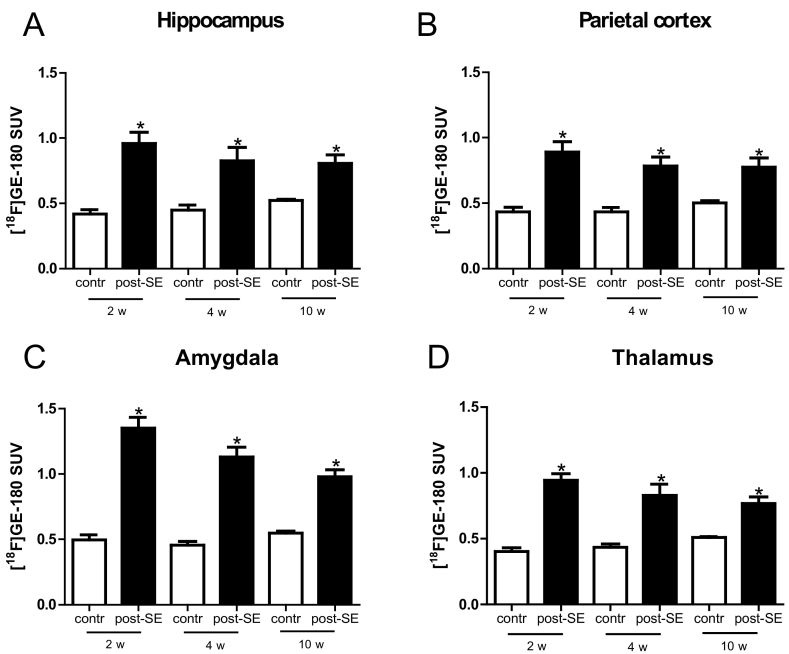
Excessive activation of inflammatory signaling pathways seems to be a hallmark of epileptogenesis. Positron emission tomography (PET) allows in vivo detection of brain inflammation with spatial information and opportunities for longitudinal follow-up scanning protocols. Here, we assessed whether molecular imaging of the 18 kDa translocator protein (TSPO) can serve as a biomarker for the development of epilepsy. Therefore, brain uptake of [F]GE-180, a highly selective radioligand of TSPO, was investigated in a longitudinal PET study in a chronic rat model of temporal lobe epilepsy. Analyses revealed that the influence of the epileptogenic insult on [F]GE-180 brain uptake was most pronounced in the earlier phase of epileptogenesis. Differences were evident in various brain regions during earlier phases of epileptogenesis with [F]GE-180 standardized uptake value enhanced by 2.1 to 2.7fold. In contrast, brain regions exhibiting differences seemed to be more restricted with less pronounced increases of tracer uptake by 1.8-2.5fold four weeks following status epilepticus and by 1.5-1.8fold in the chronic phase. Based on correlation analysis, we were able to identify regions with a predictive value showing a correlation with seizure development. These regions include the amygdala as well as a cluster of brain areas. This cluster comprises parts of different brain regions, e.g. the hippocampus, parietal cortex, thalamus, and somatosensory cortex. In conclusion, the data provide evidence that [F]GE-180 PET brain imaging can serve as a biomarker of epileptogenesis. The identification of brain regions with predictive value might facilitate the development of preventive concepts as well as the early assessment of the interventional success. Future studies are necessary to further confirm the predictivity of the approach.
炎症信号通路的过度激活似乎是癫痫发生的一个标志。正电子发射断层扫描(PET)能够在体内检测脑炎症,提供空间信息并具备纵向随访扫描方案的机会。在此,我们评估了18 kDa转运体蛋白(TSPO)的分子成像是否可作为癫痫发展的生物标志物。因此,在颞叶癫痫慢性大鼠模型的纵向PET研究中,对TSPO的高选择性放射性配体[F]GE - 180的脑摄取情况进行了研究。分析表明,致痫损伤对[F]GE - 180脑摄取的影响在癫痫发生的早期最为明显。在癫痫发生的早期阶段,不同脑区存在明显差异,[F]GE - 180标准化摄取值提高了2.1至2.7倍。相比之下,癫痫持续状态四周后,显示出差异的脑区似乎更局限,示踪剂摄取的增加不太明显,为1.8 - 2.5倍,而在慢性期为1.5 - 1.8倍。基于相关性分析,我们能够识别出与癫痫发作发展相关的具有预测价值的区域。这些区域包括杏仁核以及一组脑区。这组脑区包括不同脑区的部分区域,如海马体、顶叶皮质、丘脑和体感皮质。总之,数据表明[F]GE - 180 PET脑成像可作为癫痫发生的生物标志物。识别具有预测价值的脑区可能有助于预防概念的发展以及对干预成功与否的早期评估。未来的研究有必要进一步证实该方法的预测性。