Nabeya Daijiro, Haranaga Shusaku, Parrott Gretchen Lynn, Kinjo Takeshi, Nahar Saifun, Tanaka Teruhisa, Hirata Tetsuo, Hokama Akira, Tateyama Masao, Fujita Jiro
Department of Infectious Diseases, Respiratory, and Digestive Medicine, Graduate School of Medicine, University of the Ryukyus, 207 Uehara, Nishihara, Okinawa, 903-0215, Japan.
BMC Infect Dis. 2017 May 2;17(1):320. doi: 10.1186/s12879-017-2430-9.
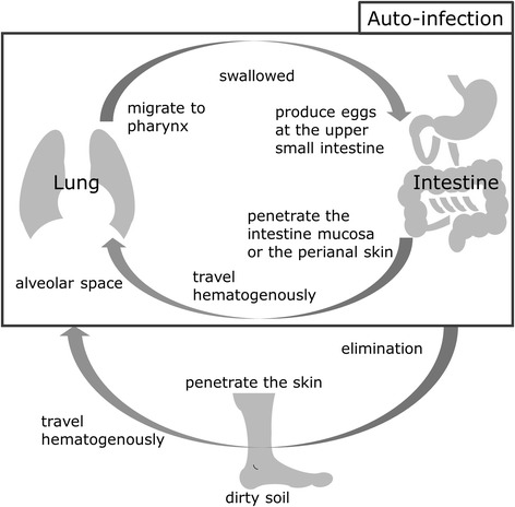
Strongyloidiasis is a chronic parasitic infection caused by Strongyloides stercoralis. Severe cases such as, hyperinfection syndrome (HS) and disseminated strongyloidiasis (DS), can involve pulmonary manifestations. These manifestations frequently aid the diagnosis of strongyloidiasis. Here, we present the pulmonary manifestations and radiological findings of severe strongyloidiasis.
From January 2004 to December 2014, all patients diagnosed with severe strongyloidiasis at the University of the Ryukyus Hospital or affiliated hospitals in Okinawa, Japan, were included in this retrospective study. All diagnoses were confirmed by the microscopic or histopathological identification of larvae. Severe strongyloidiasis was defined by the presence of any of the following: 1) the identification of S. stercoralis from extra gastrointestinal specimens, 2) sepsis, 3) meningitis, 4) acute respiratory failure, or 5) respiratory tract hemorrhage. Patients were assigned to either HS or DS. Medical records were further reviewed to extract related clinical features and radiological findings.
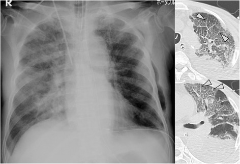
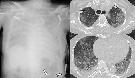
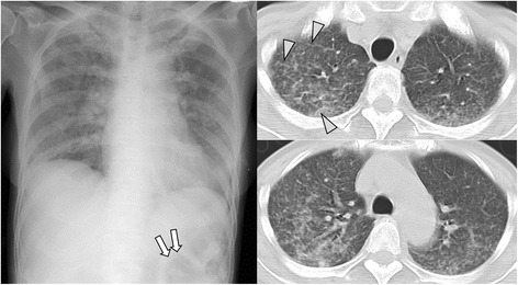
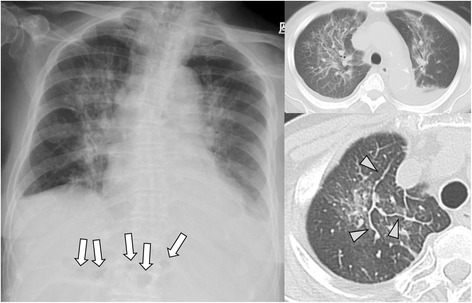
Sixteen severe strongyloidiasis cases were included. Of those, fifteen cases had pulmonary manifestations, eight had acute respiratory distress syndrome (ARDS) (53%), seven had enteric bacterial pneumonia (46%) and five had pulmonary hemorrhage (33%). Acute respiratory failure was a common indicator for pulmonary manifestation (87%). Chest X-ray findings frequently showed diffuse shadows (71%). Additionally, ileum gas was detected for ten of the sixteen cases in the upper abdomen during assessment with chest X-ray. While, chest CT findings frequently showed ground-glass opacity (GGO) in 89% of patients. Interlobular septal thickening was also frequently shown (67%), always accompanying GGO in upper lobes.
In summary, our study described HS/DS cases with pulmonary manifestations including, ARDS, bacterial pneumonia and pulmonary hemorrhage. Chest X-ray findings in HS/DS cases frequently showed diffuse shadows, and the combination of GGO and interlobular septal thickening in chest CT was common in HS/DS, regardless of accompanying pulmonary manifestations. This CT finding suggests alveolar hemorrhage could be used as a potential marker indicating the transition from latent to symptomatic state. Respiratory specimens are especially useful for detecting larvae in cases of HS/DS.
类圆线虫病是由粪类圆线虫引起的一种慢性寄生虫感染。严重病例,如超感染综合征(HS)和播散性类圆线虫病(DS),可累及肺部表现。这些表现常常有助于类圆线虫病的诊断。在此,我们呈现严重类圆线虫病的肺部表现及影像学结果。
2004年1月至2014年12月期间,所有在日本冲绳琉球大学医院或附属医院被诊断为严重类圆线虫病的患者纳入本回顾性研究。所有诊断均通过幼虫的显微镜或组织病理学鉴定得以证实。严重类圆线虫病定义为存在以下任何一种情况:1)从胃肠道外标本中鉴定出粪类圆线虫;2)败血症;3)脑膜炎;4)急性呼吸衰竭;或5)呼吸道出血。患者被分为HS或DS组。进一步查阅病历以提取相关临床特征和影像学结果。
纳入16例严重类圆线虫病病例。其中,15例有肺部表现,8例有急性呼吸窘迫综合征(ARDS)(53%),7例有肠道细菌性肺炎(46%),5例有肺出血(33%)。急性呼吸衰竭是肺部表现的常见指标(87%)。胸部X线检查结果常显示弥漫性阴影(71%)。此外,在胸部X线评估期间,16例中有10例在上腹部检测到回肠积气。而胸部CT检查结果常显示89%的患者有磨玻璃影(GGO)。小叶间隔增厚也常出现(67%),总是在上叶伴随GGO出现。
总之,我们的研究描述了伴有肺部表现的HS/DS病例,包括ARDS、细菌性肺炎和肺出血。HS/DS病例的胸部X线检查结果常显示弥漫性阴影,胸部CT中GGO和小叶间隔增厚的组合在HS/DS中很常见,无论是否伴有肺部表现。这种CT表现提示肺泡出血可作为潜在标志物,表明从潜伏状态转变为症状性状态。呼吸道标本对于检测HS/DS病例中的幼虫特别有用。