• 文献检索
  • 文档翻译
  • 深度研究
  • 学术资讯
  • Suppr Zotero 插件Zotero 插件
  • 邀请有礼
  • 套餐&价格
  • 历史记录
应用&插件
Suppr Zotero 插件Zotero 插件浏览器插件Mac 客户端Windows 客户端微信小程序
定价
高级版会员购买积分包购买API积分包
服务
文献检索文档翻译深度研究API 文档MCP 服务
关于我们
关于 Suppr公司介绍联系我们用户协议隐私条款
关注我们

Suppr 超能文献

核心技术专利:CN118964589B侵权必究
粤ICP备2023148730 号-1Suppr @ 2026

文献检索

告别复杂PubMed语法,用中文像聊天一样搜索,搜遍4000万医学文献。AI智能推荐,让科研检索更轻松。

立即免费搜索

文件翻译

保留排版,准确专业,支持PDF/Word/PPT等文件格式,支持 12+语言互译。

免费翻译文档

深度研究

AI帮你快速写综述,25分钟生成高质量综述,智能提取关键信息,辅助科研写作。

立即免费体验

18F-氟脱氧葡萄糖正电子发射断层扫描/计算机断层扫描偶然发现的一例罕见的冬眠瘤合并肺癌患者

A Rare Case of Hibernoma Occasionally Identified by 18F-fluorodeoxyglucose Positron Emission Tomography/Computed Tomography in a Patient with Lung Cancer.

作者信息

Penna Daniele, Quartuccio Natale, Testa Claudio, Arena Vincenzo, Cistaro Angelina, Pelosi Ettore

机构信息

PET/CT center, Affidea IRMET - Turin - Italy.

Wolfson Molecular Imaging Centre, University of Manchester.

出版信息

Cureus. 2017 Mar 29;9(3):e1124. doi: 10.7759/cureus.1124.

DOI:10.7759/cureus.1124
PMID:28465871
原文链接:https://pmc.ncbi.nlm.nih.gov/articles/PMC5409814/
Abstract

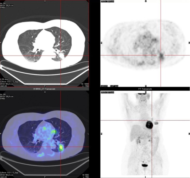
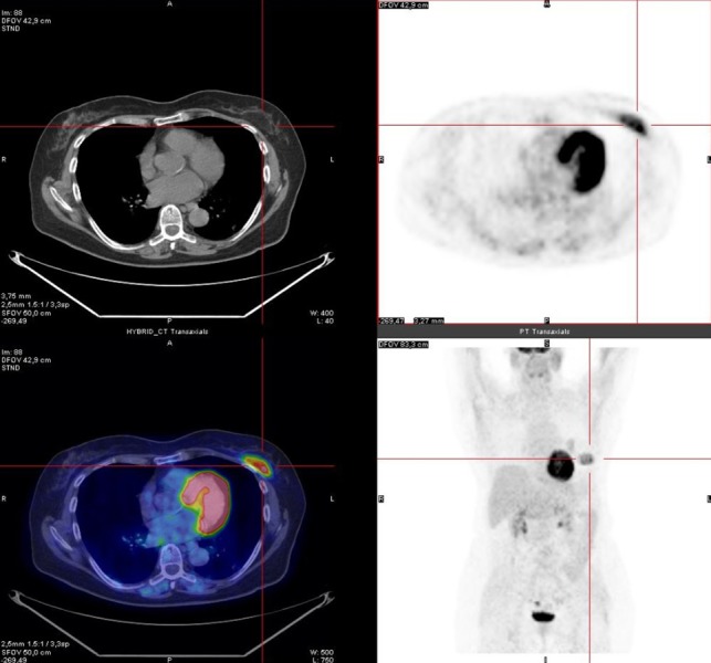
Hibernoma is a benign tumor arising from brown fat tissue. Conventional imaging techniques are not able to differentiate it from other benign lesions or malignant fatty tumors. We report the case of a 73-year-old patient who underwent a thorax computed tomography (CT) and was then referred to our department for metabolic assessment of a solitary lung nodule. An F18-fluorodeoxyglucose positron emission tomography/computed tomography (18F-FDG-PET/CT) scan was performed and demonstrated, in addition, a highly metabolic fat-containing lesion mimicking a malignant fatty tumor in the left great pectoralis muscle. The lesion was excised and resulted to be a hibernoma. This case shows that hibernoma can appear as a malignant-like lesion on 18F-FDG-PET/CT scan as per other imaging techniques, and the grade of FDG uptake does not accurately reflect malignancy in this fat-containing tumor. However, 18F-FDG-PET/CT with its whole-body scanning capability may represent a useful imaging tool in identifying, in the course of an imaging study for oncological evaluation, additional incidental findings such as benign fat-containing lesions that may require a surgical approach.

摘要

冬眠瘤是一种起源于棕色脂肪组织的良性肿瘤。传统成像技术无法将其与其他良性病变或恶性脂肪肿瘤区分开来。我们报告了一例73岁患者的病例,该患者接受了胸部计算机断层扫描(CT),随后因孤立性肺结节的代谢评估被转诊至我院。进行了一次F18-氟脱氧葡萄糖正电子发射断层扫描/计算机断层扫描(18F-FDG-PET/CT),此外还发现左胸大肌有一个高度代谢的含脂肪病变,类似恶性脂肪肿瘤。该病变被切除,结果为冬眠瘤。该病例表明,与其他成像技术一样,冬眠瘤在18F-FDG-PET/CT扫描上可表现为类似恶性的病变,并且在这种含脂肪肿瘤中,FDG摄取程度并不能准确反映恶性程度。然而,具有全身扫描能力的18F-FDG-PET/CT可能是一种有用的成像工具,在肿瘤学评估的成像研究过程中,可用于识别其他可能需要手术治疗的偶然发现,如良性含脂肪病变。

https://cdn.ncbi.nlm.nih.gov/pmc/blobs/623a/5409814/621817d9bd96/cureus-0009-00000001124-i02.jpg
https://cdn.ncbi.nlm.nih.gov/pmc/blobs/623a/5409814/57d1d65eb497/cureus-0009-00000001124-i01.jpg
https://cdn.ncbi.nlm.nih.gov/pmc/blobs/623a/5409814/621817d9bd96/cureus-0009-00000001124-i02.jpg
https://cdn.ncbi.nlm.nih.gov/pmc/blobs/623a/5409814/57d1d65eb497/cureus-0009-00000001124-i01.jpg
https://cdn.ncbi.nlm.nih.gov/pmc/blobs/623a/5409814/621817d9bd96/cureus-0009-00000001124-i02.jpg

相似文献

1
A Rare Case of Hibernoma Occasionally Identified by 18F-fluorodeoxyglucose Positron Emission Tomography/Computed Tomography in a Patient with Lung Cancer.18F-氟脱氧葡萄糖正电子发射断层扫描/计算机断层扫描偶然发现的一例罕见的冬眠瘤合并肺癌患者
Cureus. 2017 Mar 29;9(3):e1124. doi: 10.7759/cureus.1124.
2
Hibernoma: Intense Uptake on F18-FDG PET/CT.冬眠瘤:F18-FDG PET/CT 上摄取增强。
Nucl Med Mol Imaging. 2012 Sep;46(3):218-22. doi: 10.1007/s13139-012-0150-z. Epub 2012 Jun 27.
3
Hibernoma Showing High Uniform Accumulation on an F-Fluorodeoxyglucose Positron Emission Tomography Scan: A Case Report.18F-氟脱氧葡萄糖正电子发射断层扫描显示高均匀性摄取的冬眠瘤:一例报告
Case Rep Oncol. 2016 Aug 17;9(2):427-431. doi: 10.1159/000448240. eCollection 2016 May-Aug.
4
More advantages in detecting bone and soft tissue metastases from prostate cancer using F-PSMA PET/CT.使用F-PSMA PET/CT检测前列腺癌骨和软组织转移方面有更多优势。
Hell J Nucl Med. 2019 Jan-Apr;22(1):6-9. doi: 10.1967/s002449910952. Epub 2019 Mar 7.
5
False-positive findings on 18F-FDG PET/CT: differentiation of hibernoma and malignant fatty tumor on the basis of fluctuating standardized uptake values.18F-FDG PET/CT的假阳性结果:基于波动的标准化摄取值鉴别冬眠瘤与恶性脂肪肿瘤
AJR Am J Roentgenol. 2008 Apr;190(4):1091-6. doi: 10.2214/AJR.07.3061.
6
Mediastinal hibernoma simulates a malignant lesion on dual time point FDG imaging.纵隔冬眠瘤在双时相氟代脱氧葡萄糖成像上表现类似恶性病变。
Lung Cancer. 2008 Mar;59(3):391-4. doi: 10.1016/j.lungcan.2007.08.020. Epub 2007 Oct 1.
7
Case report: Intraosseous hibernoma (IOH) mimics osseous metastasis: another rare pitfall in FDG-PET-CT.病例报告:骨内冬眠瘤(IOH)酷似骨转移:18F-FDG PET-CT检查中的又一罕见陷阱
Front Nucl Med. 2023 May 17;3:1150143. doi: 10.3389/fnume.2023.1150143. eCollection 2023.
8
Dynamic (18)F-fluorodeoxyglucose positron emission tomography/CT in hibernoma: Enhanced tracer uptake mimicking liposarcoma.动态(18)F - 氟脱氧葡萄糖正电子发射断层扫描/计算机断层扫描在冬眠瘤中的应用:示踪剂摄取增加,类似脂肪肉瘤。
World J Radiol. 2013 Dec 28;5(12):498-502. doi: 10.4329/wjr.v5.i12.498.
9
Hibernoma as an incidental finding in the 18F-FDG PET/CT of a patient with melanoma.冬眠瘤在一名黑色素瘤患者的18F-FDG PET/CT检查中作为偶然发现。
Melanoma Res. 2020 Aug;30(4):420-422. doi: 10.1097/CMR.0000000000000530.
10
Propranolol 18F-FDG PET/CT: A Noninvasive Approach for Differential Diagnosis of Hibernoma and Liposarcoma.普萘洛尔 18F-FDG PET/CT:一种用于冬眠瘤和脂肪肉瘤鉴别诊断的非侵入性方法。
Clin Nucl Med. 2017 Nov;42(11):879-880. doi: 10.1097/RLU.0000000000001830.

引用本文的文献

1
A Diagnostic Dilemma of a Subcutaneous Hibernoma: Case Report.皮下脂肪瘤的诊断难题:病例报告。
Am J Case Rep. 2020 Apr 25;21:e921447. doi: 10.12659/AJCR.921447.

本文引用的文献

1
The Values and Limitations of FDG-PET/CT for Diagnosis of Hibernoma.18F-氟脱氧葡萄糖正电子发射断层显像/计算机断层扫描(FDG-PET/CT)诊断冬眠瘤的价值与局限性
Case Rep Orthop. 2015;2015:958690. doi: 10.1155/2015/958690. Epub 2015 Dec 13.
2
Hibernoma--two patients with a rare lipoid soft-tissue tumour.冬眠瘤——两名患有罕见类脂质软组织肿瘤的患者。
BMC Med Imaging. 2015 Feb 14;15:4. doi: 10.1186/s12880-015-0046-3.
3
Hibernoma: Intense Uptake on F18-FDG PET/CT.冬眠瘤:F18-FDG PET/CT 上摄取增强。
Nucl Med Mol Imaging. 2012 Sep;46(3):218-22. doi: 10.1007/s13139-012-0150-z. Epub 2012 Jun 27.
4
Intrathoracic hibernoma: a case with multimodality imaging correlation.胸腔内冬眠瘤:一例多模态影像学相关性病例。
J Thorac Imaging. 2011 Feb;26(1):W20-2. doi: 10.1097/RTI.0b013e3181e35acd.
5
Intrathoracic hibernoma: report of two cases.胸内冬眠瘤:两例报告。
Lung Cancer. 2009 Jun;64(3):367-70. doi: 10.1016/j.lungcan.2008.11.003. Epub 2009 Jan 6.
6
PET evaluation of fatty tumors in the extremity: possibility of using the standardized uptake value (SUV) to differentiate benign tumors from liposarcoma.肢体脂肪性肿瘤的正电子发射断层显像(PET)评估:利用标准化摄取值(SUV)区分良性肿瘤与脂肪肉瘤的可能性。
Ann Nucl Med. 2005 Dec;19(8):661-70. doi: 10.1007/BF02985114.
7
Patterns of (18)F-FDG uptake in adipose tissue and muscle: a potential source of false-positives for PET.脂肪组织和肌肉中(18)F-FDG摄取模式:PET假阳性的一个潜在来源。
J Nucl Med. 2003 Nov;44(11):1789-96.
8
Hibernoma of the left axilla; a case report and review of MR imaging.左腋窝冬眠瘤;病例报告及磁共振成像综述
Skeletal Radiol. 2003 May;32(5):290-4. doi: 10.1007/s00256-002-0533-9. Epub 2002 Aug 21.
9
Hibernoma: intense uptake seen on Tc-99m tetrofosmin and FDG positron emission tomographic scanning.冬眠瘤:在锝-99m 替曲膦和氟代脱氧葡萄糖正电子发射断层扫描中可见强烈摄取。
Clin Nucl Med. 2002 May;27(5):369-70. doi: 10.1097/00003072-200205000-00016.
10
The morphologic spectrum of hibernoma: a clinicopathologic study of 170 cases.冬眠瘤的形态学谱:170例临床病理研究
Am J Surg Pathol. 2001 Jun;25(6):809-14. doi: 10.1097/00000478-200106000-00014.