Mittica Gloria, Senetta Rebecca, Scotto Giulia, Aglietta Massimo, Maggiorotto Furio, Ghisoni Eleonora, Genta Sofia, Boldorini Renzo, Manini Claudia, Morra Isabella, Buosi Roberta, Sapino Anna, Cassoni Paola, Valabrega Giorgio
Department of Oncology, University of Turin, Turin, Italy.
Division of Medical Oncology-1, Candiolo Cancer Institute-FPO- IRCCS, Candiolo, Italy.
Oncotarget. 2017 Jun 20;8(25):41143-41153. doi: 10.18632/oncotarget.17068.
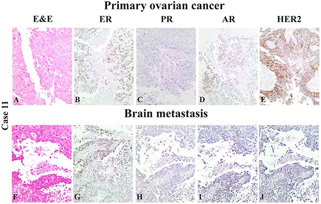
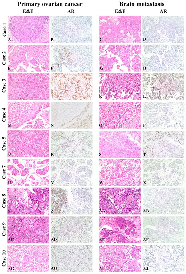
Brain metastases are uncommon localizations in epithelial ovarian cancer (EOC), their reported incidence is increasing and no predictive biomarkers have been identified yet. Goals of this study were: i) to define a possible association between Estrogen Receptor (ER), Progesterone Receptor (PR), Androgen Receptor (AR),human EGF receptor 2 (HER2) and brain progression in EOC patients, and ii) to identify differences in ER, PR, AR and HER2 protein expression from primary EOC and its matched resected brain metastasis. A retrospective series of 11 EOC with matched brain metastasis surgically removed was collected. For comparison, a "Control dataset" of 22 patients, without evidence of brain involvement after an adequate follow up was matched. ER, PR, AR and HER2 status were analyzed by means of immunohistochemistry forCases (both primary and metastatic lesions) and Controls.Univariate analysis showed that AR status was significantly associated with brain localization, both considered as discrete variable (cut-off: 10%, p=0.013) and as continuous one (p=0.035). Multivariate analysis confirmed this trend (p=0.053). When considered as continuous variables, ER and AR showed greater expression in primary tumors in comparison with brain metastases (p=0.013 and p=0.032, respectively).In our series, AR predicts brain involvement, with a 9.5 times higher propensity for AR-negative EOC. Moreover, brain dissemination is probably the result of progressive dedifferentiation of primary tumor, shown by reduction of ER and AR expression in metastases. Further studies are required, in order to anticipate and improve multimodal treatment of brain metastases.
脑转移在上皮性卵巢癌(EOC)中是不常见的转移部位,其报告发病率正在上升,且尚未发现预测性生物标志物。本研究的目的是:i)确定雌激素受体(ER)、孕激素受体(PR)、雄激素受体(AR)、人表皮生长因子受体2(HER2)与EOC患者脑转移之间的可能关联,以及ii)确定原发性EOC及其匹配的切除脑转移瘤中ER、PR、AR和HER2蛋白表达的差异。收集了11例经手术切除且伴有匹配脑转移瘤的EOC回顾性病例系列。为作比较,匹配了一个由22例患者组成的“对照数据集”,这些患者在充分随访后无脑转移证据。通过免疫组织化学分析病例组(原发性和转移性病变)和对照组的ER、PR、AR和HER2状态。单因素分析表明,AR状态与脑转移显著相关,无论是作为离散变量(临界值:10%,p=0.013)还是作为连续变量(p=0.035)。多因素分析证实了这一趋势(p=0.053)。当作为连续变量考虑时,与脑转移瘤相比,ER和AR在原发性肿瘤中的表达更高(分别为p=0.013和p=0.032)。在我们的病例系列中,AR可预测脑转移,AR阴性的EOC发生脑转移的倾向高9.5倍。此外,脑转移可能是原发性肿瘤进行性去分化的结果,转移瘤中ER和AR表达的降低表明了这一点。需要进一步研究,以预测和改善脑转移的多模式治疗。