Hayes Lawrence, Newton Charlotte
Queen Elizabeth Hospital, Kings Lynn, Norfolk, PE30 4ET, UK.
The Countess of Chester Health Park, Liverpool Road, Chester, Cheshire CH2 1UL, UK.
J Surg Case Rep. 2017 Apr 27;2017(4):rjx076. doi: 10.1093/jscr/rjx076. eCollection 2017 Apr.
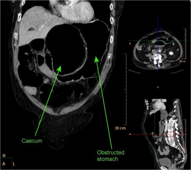
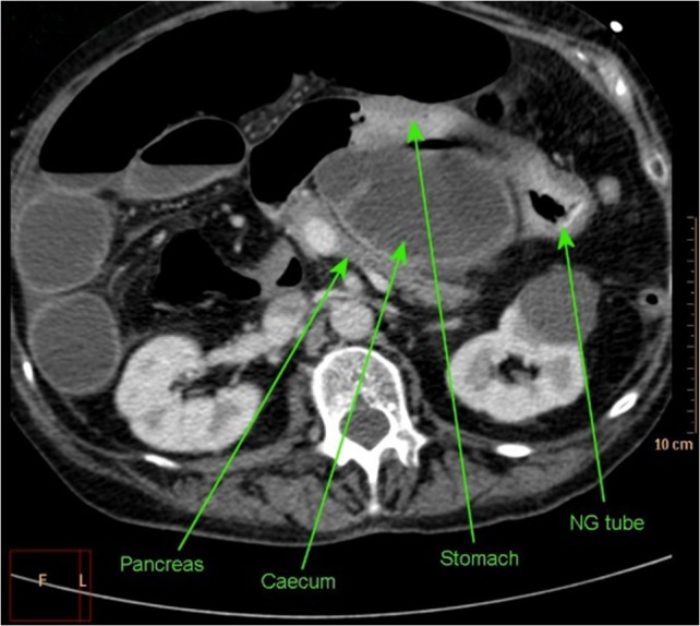
Internal hernias comprise 1% of hernias, 8% of which are through the foramen of Winslow into the lesser sac. These hernias can mimic gastric outlet obstruction and cause associated morbidity. In this case, we describe a caecal herniation into the lesser sac presenting as true gastric outlet obstruction in a 69-year-old female. Initial computed tomography (CT) imaging demonstrated a distended stomach with collapsed small bowel representing likely gastric outlet obstruction. Nasogastric tube insertion decompressed the stomach but the clinical picture progressed to that of small bowel obstruction with generalized abdominal distension and hypoactive bowel sounds. Repeat CT demonstrated caecal herniation into the lesser sac. This was confirmed at exploratory laparotomy with the caecum found in the lesser sac via the foramen of Winslow. The caecum was grossly ischaemic with patchy necrosis. A limited right hemicolectomy was performed. The patient made an uncomplicated recovery and was discharged on the eighth post-operative day.
内疝占所有疝的1%,其中8%是通过网膜孔进入小网膜囊。这些疝可类似胃出口梗阻并导致相关的发病率。在此病例中,我们描述了一名69岁女性发生盲肠疝入小网膜囊,表现为真正的胃出口梗阻。最初的计算机断层扫描(CT)成像显示胃扩张,小肠塌陷,提示可能存在胃出口梗阻。插入鼻胃管使胃减压,但临床症状进展为小肠梗阻,出现全腹膨隆和肠鸣音减弱。重复CT显示盲肠疝入小网膜囊。在剖腹探查术中证实了这一点,发现盲肠通过网膜孔位于小网膜囊内。盲肠严重缺血并有片状坏死。实施了有限的右半结肠切除术。患者恢复顺利,术后第8天出院。