Shimaoka Daisuke, Song Chenchen, Knöpfel Thomas
Neuroinformatics Japan Center (DS), RIKEN Brain Science InstituteSaitama, Japan.
Institute of Ophthalmology, University College LondonLondon, UK.
Front Cell Neurosci. 2017 Apr 24;11:108. doi: 10.3389/fncel.2017.00108. eCollection 2017.
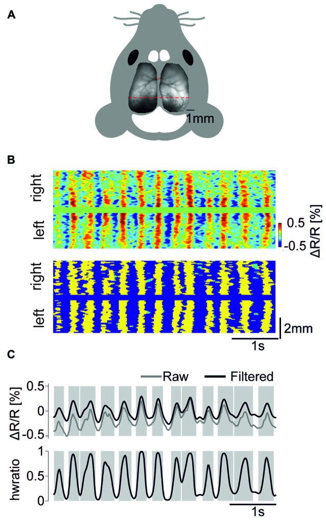
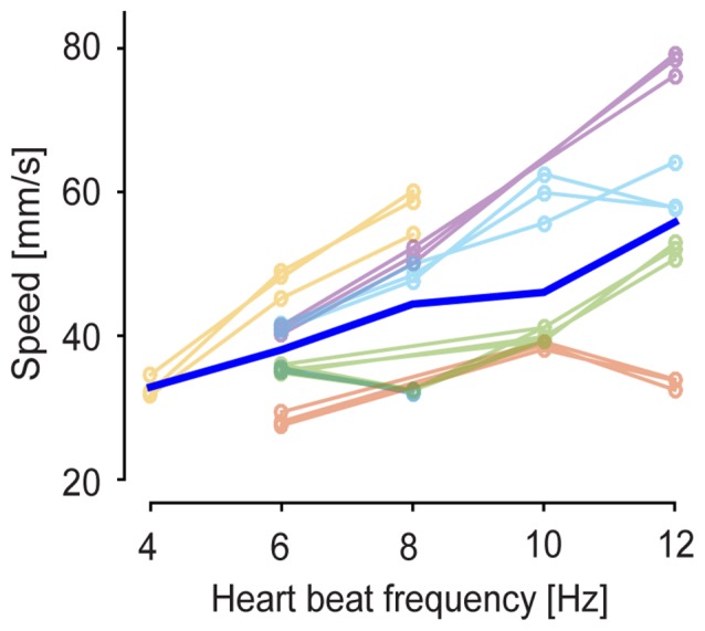
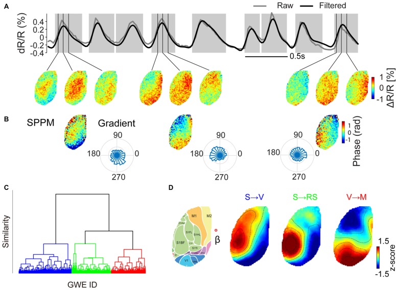
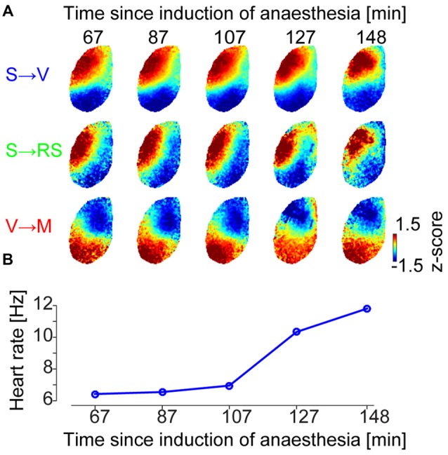
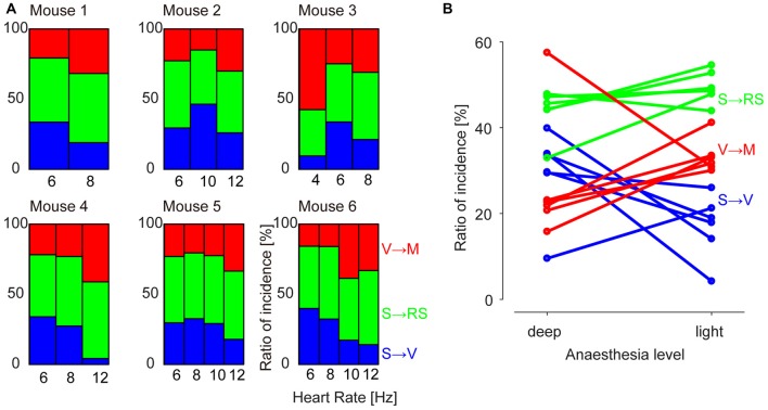
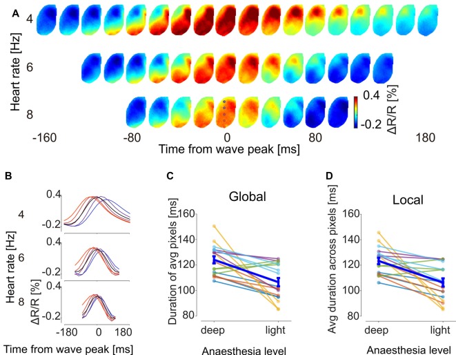
Slow cortical waves that propagate across the cerebral cortex forming large-scale spatiotemporal propagation patterns are a hallmark of non-REM sleep and anesthesia, but also occur during resting wakefulness. To investigate how the spatial temporal properties of slow waves change with the depth of anesthetic, we optically imaged population voltage transients generated by mouse layer 2/3 pyramidal neurons across one or two cortical hemispheres dorsally with a genetically encoded voltage indicator (GEVI). From deep barbiturate anesthesia to light barbiturate sedation, depolarizing wave events recruiting at least 50% of the imaged cortical area consistently appeared as a conserved repertoire of distinct wave motifs. Toward awakening, the incidence of individual motifs changed systematically (the motif propagating from visual to motor areas increased while that from somatosensory to visual areas decreased) and both local and global cortical dynamics accelerated. These findings highlight that functional endogenous interactions between distant cortical areas are not only constrained by anatomical connectivity, but can also be modulated by the brain state.
缓慢的皮层波在大脑皮层中传播,形成大规模的时空传播模式,这是非快速眼动睡眠和麻醉的一个标志,但在静息觉醒期间也会出现。为了研究慢波的时空特性如何随麻醉深度而变化,我们使用基因编码电压指示剂(GEVI)对小鼠背侧一个或两个大脑半球的2/3层锥体神经元产生的群体电压瞬变进行了光学成像。从深度巴比妥麻醉到轻度巴比妥镇静,至少招募50%成像皮层区域的去极化波事件始终表现为一组独特的波型。在接近觉醒时,各个波型的发生率会系统性地变化(从视觉区域传播到运动区域的波型增加,而从体感区域传播到视觉区域的波型减少),并且局部和整体皮层动力学都会加速。这些发现突出表明,远距离皮层区域之间的功能性内源性相互作用不仅受解剖连接性的限制,还可受脑状态的调节。