• 文献检索
  • 文档翻译
  • 深度研究
  • 学术资讯
  • Suppr Zotero 插件Zotero 插件
  • 邀请有礼
  • 套餐&价格
  • 历史记录
应用&插件
Suppr Zotero 插件Zotero 插件浏览器插件Mac 客户端Windows 客户端微信小程序
定价
高级版会员购买积分包购买API积分包
服务
文献检索文档翻译深度研究API 文档MCP 服务
关于我们
关于 Suppr公司介绍联系我们用户协议隐私条款
关注我们

Suppr 超能文献

核心技术专利:CN118964589B侵权必究
粤ICP备2023148730 号-1Suppr @ 2026

文献检索

告别复杂PubMed语法,用中文像聊天一样搜索,搜遍4000万医学文献。AI智能推荐,让科研检索更轻松。

立即免费搜索

文件翻译

保留排版,准确专业,支持PDF/Word/PPT等文件格式,支持 12+语言互译。

免费翻译文档

深度研究

AI帮你快速写综述,25分钟生成高质量综述,智能提取关键信息,辅助科研写作。

立即免费体验

直视下非穿透性 Descemet 膜剥脱性角膜内皮移植术的首次经验:病例报告

First experience of nDSAEK with heads-up surgery: A case report.

作者信息

Mohamed Yasser Helmy, Uematsu Masafumi, Inoue Daisuke, Kitaoka Takashi

机构信息

Department of Ophthalmology and Visual Sciences, Graduate School of Biomedical Sciences, Nagasaki University, Nagasaki, Japan Department of Ophthalmology, EL-Minia University Hospital, EL-Minia, Egypt.

出版信息

Medicine (Baltimore). 2017 May;96(19):e6906. doi: 10.1097/MD.0000000000006906.

DOI:10.1097/MD.0000000000006906
PMID:28489805
原文链接:https://pmc.ncbi.nlm.nih.gov/articles/PMC5428639/
Abstract

PURPOSE

To report a case of non-Descemet Stripping Automated Endothelial Keratoplasty (nDSAEK) using heads-up surgery.

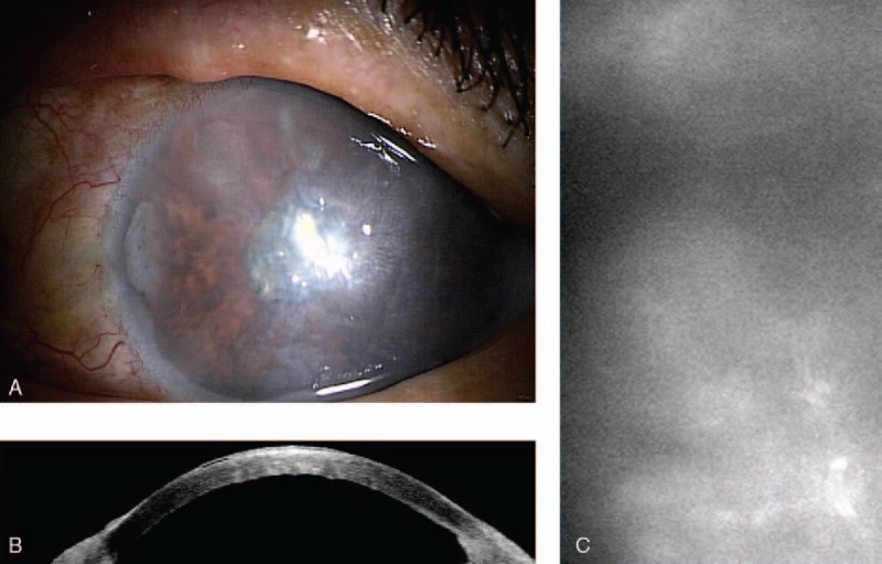
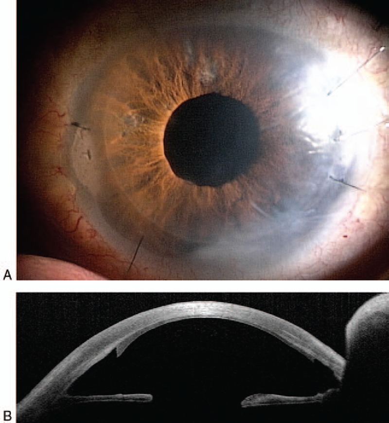
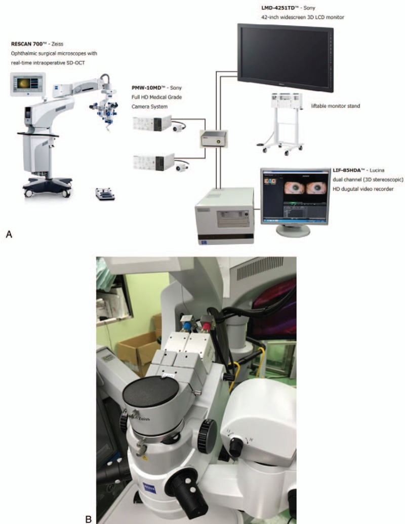
CASE/INTERVENTION: The case was a 72-years-old man who had history of left eye blunt trauma since childhood. One year ago, the patient was diagnosed to have left posttraumatic bullous keratopathy. The patient underwent lt nDSAEK by using the heads-up three-dimensional (3D) system last July. The surgery was performed with a Rescan 700 surgical microscope (Carl Zeiss), which is integrated with intraoperative optical coherence tomography (iOCT) system. During surgery, the surgeon and audience wore 3D passive polarized glasses. A 42 inch high-definition (HD) display and 2 HD cameras (Sony) were used. With this 3D system, the nDSAEK procedure before the graft insertion into the anterior chamber was easy especially with available high magnification. Also, using iOCT of the system enables the surgeon to detect any residual fluid at the donor graft-recipient interface and locate its place to be drained. The only disadvantage of the system was the difficulty in the detection of nDSAEK graft depth in the anterior chamber, which required frequent focus change during the surgery. Although the surgeon frequently adjusted the focus for clear stereoscopic view of the graft, he did not feel any eye strain or discomfort. All other steps of the procedure were performed without any problem and postoperative course of the patient was good.

CONCLUSION

Using heads-up surgery for performing anterior segment surgeries is encouraging and promising.

摘要

目的

报告一例使用抬头式手术进行非穿透性角膜内皮移植术(nDSAEK)的病例。

病例/干预措施:该病例为一名72岁男性,自童年起有左眼钝挫伤史。一年前,患者被诊断为左眼角膜外伤后大泡性角膜病变。去年7月,患者使用抬头式三维(3D)系统接受了左眼nDSAEK手术。手术使用了与术中光学相干断层扫描(iOCT)系统集成的Rescan 700手术显微镜(卡尔·蔡司公司)。手术过程中,外科医生和观众佩戴3D被动偏振眼镜。使用了一台42英寸高清(HD)显示器和两台高清摄像机(索尼公司)。借助此3D系统,在植片植入前房之前的nDSAEK手术操作轻松,尤其是在可实现高放大倍数的情况下。此外,使用该系统的iOCT可使外科医生检测供体植片与受体界面处的任何残留液体,并确定其引流位置。该系统的唯一缺点是在前房中检测nDSAEK植片深度存在困难,这在手术过程中需要频繁改变焦点。尽管外科医生频繁调整焦点以获得植片清晰的立体视图,但他并未感到任何眼疲劳或不适。手术的所有其他步骤均顺利进行,患者术后恢复良好。

结论

使用抬头式手术进行眼前节手术令人鼓舞且前景广阔。

https://cdn.ncbi.nlm.nih.gov/pmc/blobs/b3e1/5428639/09d514ee0fd3/medi-96-e6906-g004.jpg
https://cdn.ncbi.nlm.nih.gov/pmc/blobs/b3e1/5428639/6b1d5cb95eb2/medi-96-e6906-g002.jpg
https://cdn.ncbi.nlm.nih.gov/pmc/blobs/b3e1/5428639/fd5ce486b63f/medi-96-e6906-g003.jpg
https://cdn.ncbi.nlm.nih.gov/pmc/blobs/b3e1/5428639/09d514ee0fd3/medi-96-e6906-g004.jpg
https://cdn.ncbi.nlm.nih.gov/pmc/blobs/b3e1/5428639/6b1d5cb95eb2/medi-96-e6906-g002.jpg
https://cdn.ncbi.nlm.nih.gov/pmc/blobs/b3e1/5428639/fd5ce486b63f/medi-96-e6906-g003.jpg
https://cdn.ncbi.nlm.nih.gov/pmc/blobs/b3e1/5428639/09d514ee0fd3/medi-96-e6906-g004.jpg

相似文献

1
First experience of nDSAEK with heads-up surgery: A case report.直视下非穿透性 Descemet 膜剥脱性角膜内皮移植术的首次经验:病例报告
Medicine (Baltimore). 2017 May;96(19):e6906. doi: 10.1097/MD.0000000000006906.
2
Non-Descemet stripping endothelial keratoplasty for bullous keratopathy in patients with atopic dermatitis: A long-term case report.非去表皮内皮角膜移植术治疗特应性皮炎患者的大疱性角膜病变:一例长期病例报告。
Medicine (Baltimore). 2024 Sep 27;103(39):e39767. doi: 10.1097/MD.0000000000039767.
3
Intraoperative optical coherence tomography in descemet stripping automated endothelial keratoplasty: pilot experiences.Descemet膜剥除自动内皮角膜移植术中的术中光学相干断层扫描:初步经验
Int Ophthalmol. 2017 Aug;37(4):939-944. doi: 10.1007/s10792-016-0338-9. Epub 2016 Sep 21.
4
Intraoperative Interface Fluid Dynamics and Clinical Outcomes for Intraoperative Optical Coherence Tomography-Assisted Descemet Stripping Automated Endothelial Keratoplasty From the PIONEER Study.先锋研究中术中光学相干断层扫描辅助的后弹力层剥除自动角膜内皮移植术的术中界面流体动力学及临床结果
Am J Ophthalmol. 2017 Jan;173:16-22. doi: 10.1016/j.ajo.2016.09.028. Epub 2016 Oct 1.
5
[Preliminary clinical study of non-Descemet stripping endothelial keratoplasty for bullous keratopathy].非Descemet膜剥除内皮角膜移植术治疗大疱性角膜病变的初步临床研究
Zhonghua Yan Ke Za Zhi. 2010 Mar;46(3):238-42.
6
Determination of feasibility and utility of microscope-integrated optical coherence tomography during ophthalmic surgery: the DISCOVER Study RESCAN Results.眼科手术中显微镜集成光学相干断层扫描的可行性和实用性评估:DISCOVER研究RESCAN结果
JAMA Ophthalmol. 2015 Oct;133(10):1124-32. doi: 10.1001/jamaophthalmol.2015.2376.
7
Intraoperative Optical Coherence Tomography-Assisted Descemet Membrane Endothelial Keratoplasty: Toward More Efficient, Safer Surgery.术中光相干断层扫描辅助的 Descemet 膜内皮角膜移植术:追求更高效、更安全的手术。
Cornea. 2020 Jun;39(6):674-679. doi: 10.1097/ICO.0000000000002301.
8
Non-Descemet Stripping Automated Endothelial Keratoplasty for Bullous Keratopathy in Buphthalmic Eye.非后弹力层剥除自动角膜内皮移植术治疗先天性青光眼性大泡性角膜病变
Case Rep Ophthalmol. 2016 Jun 2;7(2):279-84. doi: 10.1159/000446103. eCollection 2016 May-Aug.
9
Viscoelastic-Assisted Non-Descemet Stripping Automated Endothelial Keratoplasty in Vitrectomized and Iris-Lens Diaphragm Injured Eyes.玻璃体切除及虹膜晶状体膈损伤眼中的粘弹性辅助非后弹力层剥除自动内皮角膜移植术
Eye Contact Lens. 2015 Nov;41(6):398-402. doi: 10.1097/ICL.0000000000000156.
10
Case series and techniques of Descemet's Stripping Automated Endothelial Keratoplasty for severe bullous keratopathy after birth injury.出生损伤后严重大泡性角膜病变的后弹力层剥除自动内皮角膜移植术的病例系列及技术
BMC Ophthalmol. 2015 Aug 6;15:92. doi: 10.1186/s12886-015-0094-z.

引用本文的文献

1
Non-Descemet Stripping Automated Endothelial Keratoplasty (nDSAEK) for Late Endothelial Failure After Mushroom Keratoplasty: A Retrospective Analysis of Visual and Anatomical Outcomes.非后弹力层剥除自动角膜内皮移植术(nDSAEK)治疗真菌性角膜移植术后晚期角膜内皮功能衰竭:视觉和解剖学结果的回顾性分析
J Clin Med. 2025 Aug 7;14(15):5568. doi: 10.3390/jcm14155568.
2
Use of a Novel Three-dimensional Head-mounted Digital Visualization Platform in Corneal Endothelial Transplantation.一种新型三维头戴式数字可视化平台在角膜内皮移植中的应用。
Ophthalmol Ther. 2023 Feb;12(1):625-631. doi: 10.1007/s40123-022-00624-6. Epub 2022 Dec 12.
3

本文引用的文献

1
"HEADS UP" DIGITALLY ASSISTED SURGICAL VIEWING FOR RETINAL DETACHMENT REPAIR IN A PATIENT WITH SEVERE KYPHOSIS.“抬头”数字辅助手术视野用于重度脊柱后凸患者的视网膜脱离修复
Retin Cases Brief Rep. 2018;12(3):257-259. doi: 10.1097/ICB.0000000000000486.
2
Heads-up 3D Microscopy: An Ergonomic and Educational Approach to Microsurgery.抬头式3D显微镜:一种符合人体工程学且用于显微外科手术教学的方法。
Plast Reconstr Surg Glob Open. 2016 May 25;4(5):e717. doi: 10.1097/GOX.0000000000000727. eCollection 2016 May.
3
HEADS-UP SURGERY FOR VITREORETINAL PROCEDURES: An Experimental and Clinical Study.
3D Visualization System in Descemet Membrane Endothelial Keratoplasty (DMEK): A Six-Month Comparison with Conventional Microscope.
Descemet膜内皮角膜移植术(DMEK)中的3D可视化系统:与传统显微镜的六个月比较
J Clin Med. 2022 Jul 25;11(15):4312. doi: 10.3390/jcm11154312.
4
Optimization of surgeon ergonomics with three-dimensional heads-up display for ophthalmic surgeries.三维抬头显示器优化眼科手术中的外科医生工效学。
Indian J Ophthalmol. 2022 Mar;70(3):847-850. doi: 10.4103/ijo.IJO_1548_21.
5
Digitally assisted three-dimensional surgery - Beyond vitreous.数字化辅助三维手术——超越玻璃体。
Indian J Ophthalmol. 2021 Jul;69(7):1793-1800. doi: 10.4103/ijo.IJO_3111_20.
6
Three-dimensional heads up display in anterior segment surgeries- Expanding frontiers in the COVID-19 era.眼前节手术中的三维抬头显示器——新冠疫情时代的前沿拓展
Indian J Ophthalmol. 2020 Nov;68(11):2338-2340. doi: 10.4103/ijo.IJO_2978_20.
7
Quantitative evaluation of digital-image enhancement during heads-up surgery.头高脚低位手术中数字图像增强的定量评估。
Sci Rep. 2019 Nov 4;9(1):15931. doi: 10.1038/s41598-019-52492-z.
8
First experience of nDASEK with heads-up Surgery: A case report: Erratum.使用抬头式手术进行非穿透性深板层角膜内皮移植术的首次经验:病例报告:勘误
Medicine (Baltimore). 2018 Aug;97(35):e12287. doi: 10.1097/MD.0000000000012287.
9
Heads up Descemet membrane endothelial keratoplasty performed using a 3D visualization system.使用三维可视化系统进行的前弹力层下内皮角膜移植术。
J Surg Case Rep. 2017 Nov 28;2017(11):rjx231. doi: 10.1093/jscr/rjx231. eCollection 2017 Nov.
玻璃体视网膜手术的术中直视:一项实验与临床研究。
Retina. 2016 Jan;36(1):137-47. doi: 10.1097/IAE.0000000000000689.
4
Integrative advances for OCT-guided ophthalmic surgery and intraoperative OCT: microscope integration, surgical instrumentation, and heads-up display surgeon feedback.光学相干断层扫描(OCT)引导的眼科手术及术中OCT的综合进展:显微镜集成、手术器械及平视显示器外科医生反馈
PLoS One. 2014 Aug 20;9(8):e105224. doi: 10.1371/journal.pone.0105224. eCollection 2014.
5
Occupational injury in plastic surgeons.整形医师职业伤害。
Plast Reconstr Surg. 2010 May;125(5):1555-1561. doi: 10.1097/PRS.0b013e3181d62a94.
6
Non-Descemet stripping automated endothelial keratoplasty for endothelial dysfunction secondary to argon laser iridotomy.非Descemet剥除自动内皮角膜移植术治疗氩激光虹膜切开术后继发的内皮功能障碍。
Am J Ophthalmol. 2008 Oct;146(4):543-549. doi: 10.1016/j.ajo.2008.05.028. Epub 2008 Jul 9.
7
Evaluation of a stereoscopic camera-based three-dimensional viewing workstation for ophthalmic surgery.基于立体相机的眼科手术三维观察工作站的评估
Am J Ophthalmol. 2007 May;143(5):891-2. doi: 10.1016/j.ajo.2006.12.032.
8
Symptoms of musculoskeletal disorders in ophthalmologists.眼科医生的肌肉骨骼疾病症状。
Am J Ophthalmol. 2005 Jan;139(1):179-81. doi: 10.1016/j.ajo.2004.06.091.
9
The 45 degree tilt: improvement in surgical ergonomics.45度倾斜:手术人体工程学的改善。
J Cataract Refract Surg. 1999 Feb;25(2):174-6. doi: 10.1016/s0886-3350(99)80122-9.