Rabiolo Alessandro, Parravano Mariacristina, Querques Lea, Cicinelli Maria Vittoria, Carnevali Adriano, Sacconi Riccardo, Centoducati Teresa, Vujosevic Stela, Bandello Francesco, Querques Giuseppe
Department of Ophthalmology, University Vita-Salute, Scientific Institute San Raffaele, Milan.
G. B. Bietti Foundation - IRCCS, Rome.
Clin Ophthalmol. 2017 Apr 27;11:803-807. doi: 10.2147/OPTH.S133637. eCollection 2017.
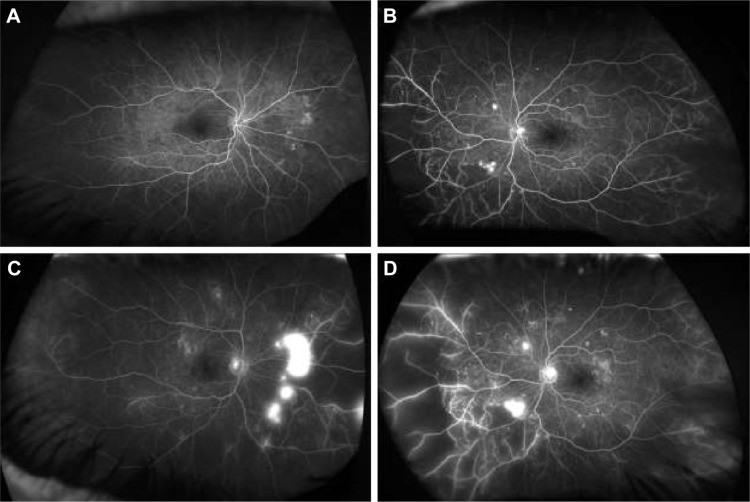
Fluorescein angiography (FA) is a useful examination in patients suffering from diabetic retinopathy (DR). Traditional angiograms explore 30°-50° of the retina at once; however, visualization of peripheral retina is fundamental in order to assess nonperfused areas, vascular leakage, microvascular abnormalities, and neovascularizations. In order to expand the field of view, wide-field and ultra-wide-field imaging has been developed allowing to image up to 200° of retinal surface in one single shot. The aim of this narrative review was to provide an overview of the role of the most recent technique of ultra-wide-field fluorescein angiography in DR.
荧光素血管造影(FA)对糖尿病视网膜病变(DR)患者是一种有用的检查方法。传统血管造影一次只能探查视网膜30°-50°的范围;然而,为了评估无灌注区、血管渗漏、微血管异常和新生血管,视网膜周边部的显像是至关重要的。为了扩大视野,已经开发出广角和超广角成像技术,能够单次成像多达200°的视网膜表面。本叙述性综述的目的是概述超广角荧光素血管造影这项最新技术在DR中的作用。