Taroni Mathieu, Seurin Marie-Jose, Carozzo Claude, Escriou Catherine
VetAgro Sup, Lyon, France.
CIRMA, Lyon, France.
JFMS Open Rep. 2015 Jul 14;1(2):2055116915593970. doi: 10.1177/2055116915593970. eCollection 2015 Jul-Dec.
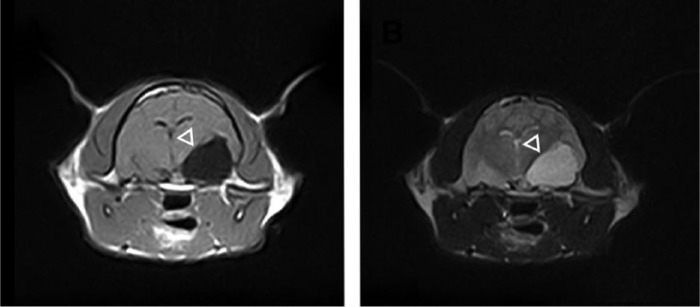
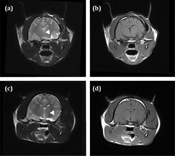
Arachnoid cysts are defined as an accumulation of fluid within the arachnoid membrane. Feline intracranial arachnoid cysts are seldom reported, with only three cases in the veterinary literature. A 1-year-old male neutered European cat with a 24 h history of seizures was presented to the small animal neurology department at Vetagro Sup, Lyon. Magnetic resonance imaging (MRI) revealed a large intracranial arachnoid cyst ventral to the brain in the left temporal area. Cystoperitoneal shunt placement resulted in complete resolution of the cyst without recurrence (follow-up MRIs 3 weeks and 21 months after surgery). Anticonvulsant treatment (phenobarbital 2.5 mg/kg q12h) was initiated at presentation and gradually stopped after 17 months. Seizures recurred 4 months after ending treatment, and seizure therapy was therefore restarted at the initial dose. We report a case of an intracranial arachnoid cyst in an unusual location not previously described. A cystoperitoneal shunt resolved the cyst without complications. Maintenance anticonvulsant treatment was required to control symptomatic epilepsy.
蛛网膜囊肿被定义为蛛网膜膜内的液体聚集。猫颅内蛛网膜囊肿鲜有报道,兽医文献中仅有三例。一只1岁绝育的欧洲雄性猫,有24小时癫痫发作史,被送至里昂维塔格罗苏普小动物神经科。磁共振成像(MRI)显示左颞区脑腹侧有一个大的颅内蛛网膜囊肿。囊肿-腹腔分流术使囊肿完全消退且无复发(术后3周和21个月的随访MRI)。就诊时开始抗惊厥治疗(苯巴比妥2.5mg/kg,每12小时一次),17个月后逐渐停药。停药4个月后癫痫复发,因此以初始剂量重新开始癫痫治疗。我们报告了一例位于异常位置的颅内蛛网膜囊肿病例,该位置此前未被描述过。囊肿-腹腔分流术使囊肿消退且无并发症。需要维持抗惊厥治疗以控制症状性癫痫。