Asano Shuichi, Ito Satoru, Takahashi Kota, Furuya Kishio, Kondo Masashi, Sokabe Masahiro, Hasegawa Yoshinori
Department of Respiratory Medicine, Nagoya University Graduate School of Medicine, Nagoya, Japan.
Department of Respiratory Medicine, Nagoya University Graduate School of Medicine, Nagoya, Japan
Physiol Rep. 2017 May;5(9). doi: 10.14814/phy2.13281. Epub 2017 May 14.
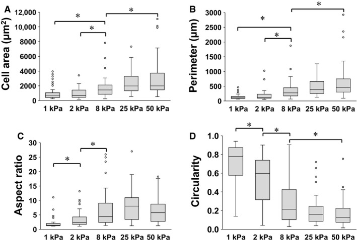
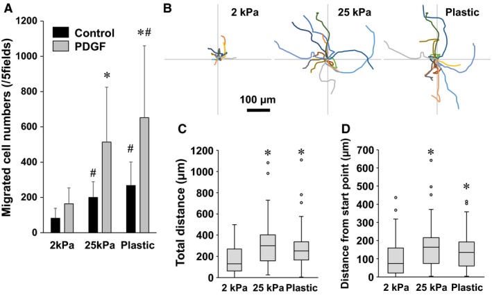
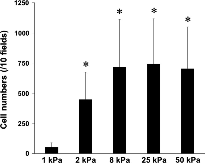
In patients with pulmonary diseases such as idiopathic pulmonary fibrosis and severe acute respiratory distress syndrome, progressive pulmonary fibrosis is caused by dysregulated wound healing via activation of fibroblasts after lung inflammation or severe damage. Migration of fibroblasts toward the fibrotic lesions plays an important role in pulmonary fibrosis. Fibrotic tissue in the lung is much stiffer than normal lung tissue. Emerging evidence supports the hypothesis that the stiffness of the matrix is not only a consequence of fibrosis, but also can induce fibroblast activation. Nevertheless, the effects of substrate rigidity on migration of lung fibroblasts have not been fully elucidated. We evaluated the effects of substrate stiffness on the morphology, -smooth muscle actin (-SMA) expression, and cell migration of primary human lung fibroblasts by using polyacrylamide hydrogels with stiffnesses ranging from 1 to 50 kPa. Cell motility was assessed by platelet-derived growth factor (PDGF)-induced chemotaxis and random walk migration assays. As the stiffness of substrates increased, fibroblasts became spindle-shaped and spread. Expression of -SMA proteins was higher on the stiffer substrates (25 kPa gel and plastic dishes) than on the soft 2 kPa gel. Both PDGF-induced chemotaxis and random walk migration of fibroblasts precultured on stiff substrates (25 kPa gel and plastic dishes) were significantly higher than those of cells precultured on 2 kPa gel. Transfection of the fibroblasts with short interfering RNA for -SMA inhibited cell migration. These findings suggest that fibroblast activation induced by a stiff matrix is involved in mechanisms of the pathophysiology of pulmonary fibrosis.
在患有诸如特发性肺纤维化和严重急性呼吸窘迫综合征等肺部疾病的患者中,进行性肺纤维化是由肺部炎症或严重损伤后成纤维细胞激活导致的伤口愈合失调所引起的。成纤维细胞向纤维化病灶的迁移在肺纤维化中起重要作用。肺中的纤维化组织比正常肺组织硬得多。新出现的证据支持这样一种假说,即基质的硬度不仅是纤维化的结果,而且还可诱导成纤维细胞激活。然而,底物硬度对肺成纤维细胞迁移的影响尚未完全阐明。我们使用刚度范围为1至50 kPa的聚丙烯酰胺水凝胶评估了底物硬度对原代人肺成纤维细胞的形态、α-平滑肌肌动蛋白(α-SMA)表达和细胞迁移的影响。通过血小板衍生生长因子(PDGF)诱导的趋化作用和随机游走迁移试验评估细胞运动性。随着底物硬度的增加,成纤维细胞变成纺锤形并伸展。在较硬的底物(25 kPa凝胶和塑料培养皿)上,α-SMA蛋白的表达高于在柔软的2 kPa凝胶上。在硬底物(25 kPa凝胶和塑料培养皿)上预培养的成纤维细胞的PDGF诱导趋化作用和随机游走迁移均明显高于在2 kPa凝胶上预培养的细胞。用针对α-SMA的短干扰RNA转染成纤维细胞可抑制细胞迁移。这些发现表明,硬基质诱导的成纤维细胞激活参与了肺纤维化病理生理学机制。