Gong Jianhua, Wang Junyi, Tian Yu, Zhang Jing, Liang Wenjin, Li Zeming, Yu Jidong, Tang Bo, He Songqing
Department of Hepatobiliary Surgery and Laboratory, Affiliated Hospital of Guilin Medical University, Guilin, Guangxi 541001, P.R. China.
Division of Hepatobiliary and Pancreatic Surgery, Department of Surgery, The Second Hospital of Dalian Medical University, Dalian, Liaoning 116027, P.R. China.
Biomed Rep. 2017 May;6(5):525-531. doi: 10.3892/br.2017.891. Epub 2017 Apr 11.
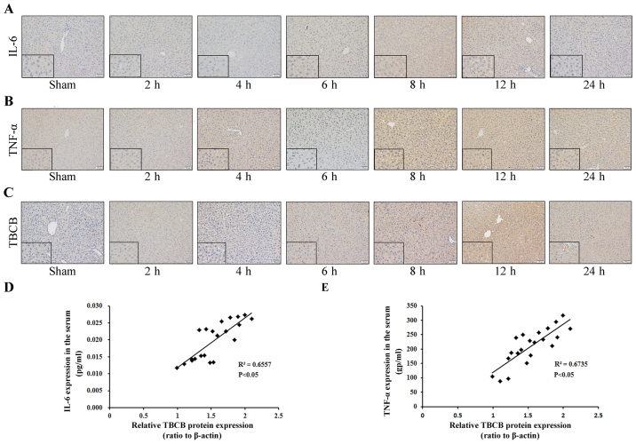
The aim of the present study was to investigate the association between tubulin folding cofactor B (TBCB) expression and ischemia-reperfusion injury (IRI) in mice. A total of 48 C57BL/6 mice were randomly divided into a control group (Sham, n=6) and an ischemia-reperfusion group (n=42). The ischemia-reperfusion group was further divided into 6 subgroups as per different times after reperfusion (2, 4, 6, 8, 12 and 24 h), with 7 mice per subgroup. A hepatic IRI model was established in mice by clamping the hepatic hilum. Morphology, serum levels of alanine aminotransferase (ALT), aspartate aminotransferase (AST), interleukin 6 (IL-6) and tumor necrosis factor-α (TNF-α), and the expression level of TBCB were detected. Compared with the control group, the livers from the ischemia-reperfusion group were significantly changed, particularly at 12 h following ischemia-reperfusion, with obvious hepatic cell degeneration and necrosis. The ALT, AST, IL-6 and TNF-α levels in the sera of the mice in the hepatic ischemia-reperfusion group were increased at all time points following ischemia-reperfusion, and were the highest at 12 h, demonstrating statistically significant differences when compared with the control group (P<0.05). Furthermore, the expression levels of TBCB, TNF-α and IL-6 were significantly increased at all time-points following ischemia-reperfusion, and were the most significant at 12 h. At 24 h following ischemia-reperfusion, the expression levels had decreased. The present study indicated that TBCB expression is associated with TNF-α and IL-6 expression levels in mice with hepatic ischemia-reperfusion, and may be key in the development of liver injury during ischemia-reperfusion in mice.
本研究的目的是探讨微管蛋白折叠辅助因子B(TBCB)表达与小鼠缺血再灌注损伤(IRI)之间的关联。总共48只C57BL/6小鼠被随机分为对照组(假手术组,n = 6)和缺血再灌注组(n = 42)。缺血再灌注组根据再灌注后的不同时间(2、4、6、8、12和24小时)进一步分为6个亚组,每组7只小鼠。通过夹闭肝门在小鼠中建立肝IRI模型。检测形态学、血清丙氨酸氨基转移酶(ALT)、天冬氨酸氨基转移酶(AST)、白细胞介素6(IL-6)和肿瘤坏死因子-α(TNF-α)水平以及TBCB的表达水平。与对照组相比,缺血再灌注组的肝脏有明显变化,特别是在缺血再灌注后12小时,肝细胞有明显的变性和坏死。肝缺血再灌注组小鼠血清中的ALT、AST、IL-6和TNF-α水平在缺血再灌注后的所有时间点均升高,且在12小时时最高,与对照组相比差异有统计学意义(P<0.05)。此外,缺血再灌注后所有时间点TBCB、TNF-α和IL-6的表达水平均显著升高,且在12小时时最为显著。缺血再灌注后24小时,表达水平有所下降。本研究表明,TBCB表达与肝缺血再灌注小鼠的TNF-α和IL-6表达水平相关,可能是小鼠缺血再灌注期间肝损伤发生的关键因素。