Chao Jennifer R, Lamba Deepak A, Klesert Todd R, Torre Anna La, Hoshino Akina, Taylor Russell J, Jayabalu Anu, Engel Abbi L, Khuu Thomas H, Wang Ruikang K, Neitz Maureen, Neitz Jay, Reh Thomas A
Department of Ophthalmology, University of Washington, Seattle, WA, USA.
Buck Institute for Research on Aging, Novato, CA, USA.
Transl Vis Sci Technol. 2017 May 16;6(3):4. doi: 10.1167/tvst.6.3.4. eCollection 2017 May.
Previous studies have demonstrated the ability of retinal cells derived from human embryonic stem cells (hESCs) to survive, integrate into the host retina, and mediate light responses in murine mouse models. Our aim is to determine whether these cells can also survive and integrate into the retina of a nonhuman primate, following transplantation into the subretinal space.
hESCs were differentiated toward retinal neuronal fates using our previously published technique and cultured for 60 to 70 days. Differentiated cells were further treated with 20 μM N-[N-(3,5-Difluorophenacetyl)-L-alanyl]-S-phenylglycine t-butyl ester (DAPT) for a period of 5 days immediately prior to subretinal transplantation. Differentiated cells were labeled with a lentivirus expressing GFP. One million cells (10,000 cells/μL) were injected into the submacular space into a squirrel monkey eye, using an ab externo technique.
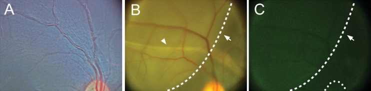
RetCam imaging demonstrated the presence and survival of human donor cells 3 months after transplantation in the eye. Injected cells consolidated in the temporal macula. GFP axonal projections were observed to emanate from the central consolidation of cells at 1 month, with some projecting into the optic nerve by 3 months after transplantation.
Human ES cell-derived retinal neurons injected into the submacular space of a squirrel monkey survive at least 3 months postinjection without immunosuppression. Some donor cells appeared to integrate into the host inner retina, and numerous donor axonal projections were noted throughout, with some projecting into the optic nerve.
These data illustrate the feasibility of hESC-derived retinal cell replacement in the nonhuman primate eye.
先前的研究已经证明,源自人类胚胎干细胞(hESCs)的视网膜细胞能够在小鼠模型中存活、整合到宿主视网膜中并介导光反应。我们的目的是确定这些细胞在移植到视网膜下间隙后,是否也能在非人灵长类动物的视网膜中存活并整合。
使用我们先前发表的技术将hESCs诱导分化为视网膜神经元命运,并培养60至70天。在视网膜下移植前,将分化的细胞用20μM N-[N-(3,5-二氟苯乙酰基)-L-丙氨酰基]-S-苯甘氨酸叔丁酯(DAPT)进一步处理5天。用表达绿色荧光蛋白(GFP)的慢病毒标记分化的细胞。采用经外路技术,将100万个细胞(10,000个细胞/μL)注入松鼠猴眼的黄斑下间隙。
RetCam成像显示移植后3个月人供体细胞在眼中的存在和存活情况。注入的细胞在颞侧黄斑处聚集。移植后1个月观察到GFP轴突投射从细胞的中央聚集处发出,到3个月时一些投射到视神经中。
注入松鼠猴黄斑下间隙的人胚胎干细胞来源的视网膜神经元在注射后至少存活3个月,无需免疫抑制。一些供体细胞似乎整合到宿主的内视网膜中,并且在整个区域都观察到大量的供体轴突投射,其中一些投射到视神经中。
这些数据说明了人胚胎干细胞来源的视网膜细胞替代在非人灵长类动物眼中的可行性。