Ji Meiying, Jin Aihua, Sun Jie, Cui Xuelian, Yang Yang, Chen Liyan, Lin Zhenhua
Cancer Research Center and Department of Pathology, Yanbian University Medical College, Yanji, Jilin 133002, P.R. China.
Department of Internal Medicine, Yanbian University Hospital, Yanji, Jilin 133000, P.R. China.
Oncol Lett. 2017 May;13(5):2996-3002. doi: 10.3892/ol.2017.5821. Epub 2017 Mar 7.
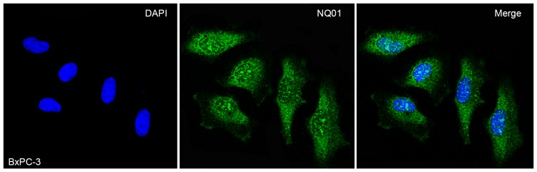
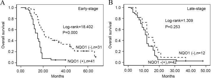
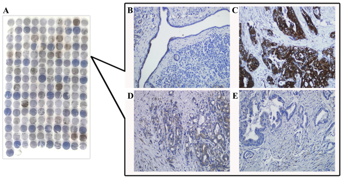
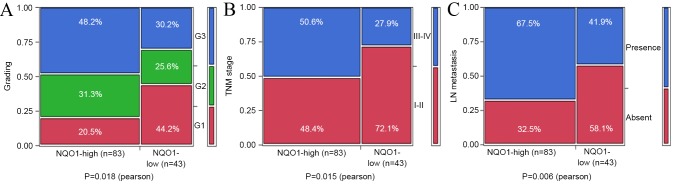
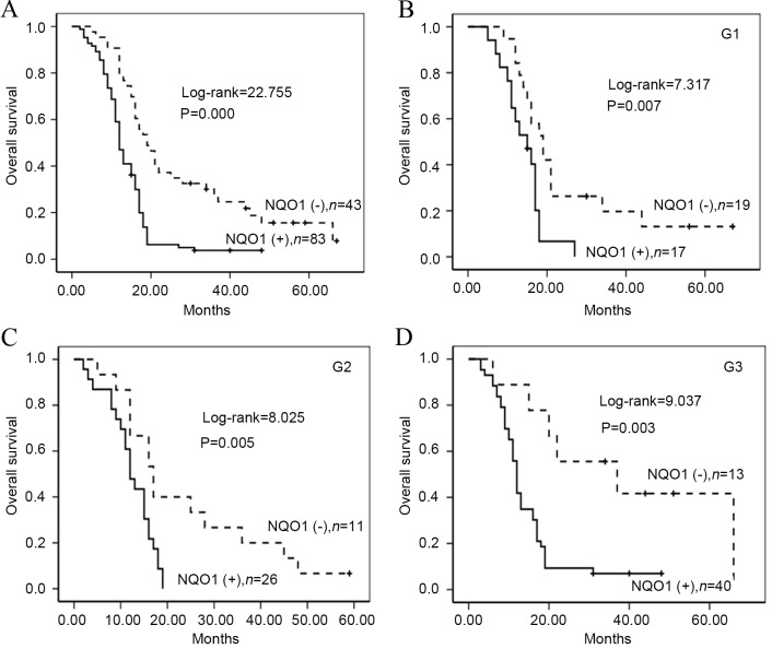
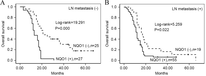
Nicotinamide adenine dinucleotide phosphate: quinone oxidoreductase 1 (NQO1) protects cells from oxidative damage. NQO1 has been reported to be upregulated in numerous solid tumors, suggesting a role in carcinogenesis and tumor progression. The present study attempted to investigate the clinical prognostic significance of NQO1 overexpression in pancreatic ductal adenocarcinoma (PDAC). A total of 181 tissue specimens were studied, including 126 PDAC and 55 normal pancreas specimens, which were selected for immunohistochemical staining of NQO1 protein. Immunofluorescence staining was additionally performed to identify the subcellular localization of NQO1 protein in pancreatic cancer BxPC-3 cells. The association between NQO1 overexpression and the clinical features of PDAC were evaluated by χ and Fisher's exact test. Overall survival of PDAC patients was calculated using Kaplan-Meier analysis. Univariate and multivariate analyses were performed using the Cox proportional hazards regression model. The NQO1 protein was mainly located in the cytoplasm and nucleus of BxPC-3 cells. The strongly positive rate of NQO1 expression in PDAC (65.9%, 83/126) was increased compared with that in normal pancreatic tissues (10.9%, 6/55). The positive rate of NQO1 protein was associated with grading, lymph node stage and tumor-node-metastasis (TNM) stage. Additionally, multivariate analysis suggested that NQO1 was a significant independent prognostic factor along with TNM stage in PDAC. In conclusion, high expression of NQO1 appears to be associated with PDAC progression, and may be an independent prognostic biomarker in PDAC.
烟酰胺腺嘌呤二核苷酸磷酸醌氧化还原酶1(NQO1)可保护细胞免受氧化损伤。据报道,NQO1在多种实体瘤中表达上调,提示其在致癌作用和肿瘤进展中发挥作用。本研究旨在探讨NQO1过表达在胰腺导管腺癌(PDAC)中的临床预后意义。共研究了181份组织标本,包括126份PDAC标本和55份正常胰腺标本,选取这些标本进行NQO1蛋白的免疫组织化学染色。另外进行免疫荧光染色以确定NQO1蛋白在胰腺癌BxPC-3细胞中的亚细胞定位。通过χ检验和Fisher精确检验评估NQO1过表达与PDAC临床特征之间的关联。采用Kaplan-Meier分析计算PDAC患者的总生存期。使用Cox比例风险回归模型进行单因素和多因素分析。NQO1蛋白主要位于BxPC-3细胞的细胞质和细胞核中。与正常胰腺组织(10.9%,6/55)相比,PDAC中NQO1表达的强阳性率升高(65.9%,83/126)。NQO1蛋白的阳性率与分级、淋巴结分期和肿瘤-淋巴结-转移(TNM)分期相关。此外,多因素分析表明,在PDAC中,NQO1与TNM分期一样是一个重要的独立预后因素。总之,NQO1的高表达似乎与PDAC进展相关,并且可能是PDAC中的一个独立预后生物标志物。