• 文献检索
  • 文档翻译
  • 深度研究
  • 学术资讯
  • Suppr Zotero 插件Zotero 插件
  • 邀请有礼
  • 套餐&价格
  • 历史记录
应用&插件
Suppr Zotero 插件Zotero 插件浏览器插件Mac 客户端Windows 客户端微信小程序
定价
高级版会员购买积分包购买API积分包
服务
文献检索文档翻译深度研究API 文档MCP 服务
关于我们
关于 Suppr公司介绍联系我们用户协议隐私条款
关注我们

Suppr 超能文献

核心技术专利:CN118964589B侵权必究
粤ICP备2023148730 号-1Suppr @ 2026

文献检索

告别复杂PubMed语法,用中文像聊天一样搜索,搜遍4000万医学文献。AI智能推荐,让科研检索更轻松。

立即免费搜索

文件翻译

保留排版,准确专业,支持PDF/Word/PPT等文件格式,支持 12+语言互译。

免费翻译文档

深度研究

AI帮你快速写综述,25分钟生成高质量综述,智能提取关键信息,辅助科研写作。

立即免费体验

口腔癌细胞上尿激酶受体(uPAR)的裂解:转化生长因子-β1(TGF-β1)的调节作用及其对迁移和侵袭的潜在影响

Cleavage of the urokinase receptor (uPAR) on oral cancer cells: regulation by transforming growth factor - β1 (TGF-β1) and potential effects on migration and invasion.

作者信息

Magnussen Synnove Norvoll, Hadler-Olsen Elin, Costea Daniela Elena, Berg Eli, Jacobsen Cristiane Cavalcanti, Mortensen Bente, Salo Tuula, Martinez-Zubiaurre Inigo, Winberg Jan-Olof, Uhlin-Hansen Lars, Svineng Gunbjorg

机构信息

Department of Medical Biology, Faculty of Health Sciences, UiT - The Arctic University of Norway, N-9037, Tromsø, Norway.

Diagnostic Clinic - Clinical Pathology, University Hospital of North Norway, Tromsø, Norway.

出版信息

BMC Cancer. 2017 May 19;17(1):350. doi: 10.1186/s12885-017-3349-7.

DOI:10.1186/s12885-017-3349-7
PMID:28526008
原文链接:https://pmc.ncbi.nlm.nih.gov/articles/PMC5438506/
Abstract

BACKGROUND

Urokinase plasminogen activator (uPA) receptor (uPAR) is up-regulated at the invasive tumour front of human oral squamous cell carcinoma (OSCC), indicating a role for uPAR in tumour progression. We previously observed elevated expression of uPAR at the tumour-stroma interface in a mouse model for OSCC, which was associated with increased proteolytic activity. The tumour microenvironment regulated uPAR expression, as well as its glycosylation and cleavage. Both full-length- and cleaved uPAR (uPAR (II-III)) are involved in highly regulated processes such as cell signalling, proliferation, migration, stem cell mobilization and invasion. The aim of the current study was to analyse tumour associated factors and their effect on uPAR cleavage, and the potential implications for cell proliferation, migration and invasion.

METHODS

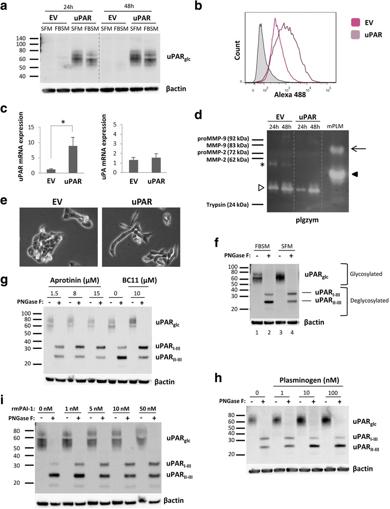
Mouse uPAR was stably overexpressed in the mouse OSCC cell line AT84. The ratio of full-length versus cleaved uPAR as analysed by Western blotting and its regulation was assessed by addition of different protease inhibitors and transforming growth factor - β1 (TGF-β1). The role of uPAR cleavage in cell proliferation and migration was analysed using real-time cell analysis and invasion was assessed using the myoma invasion model.

RESULTS

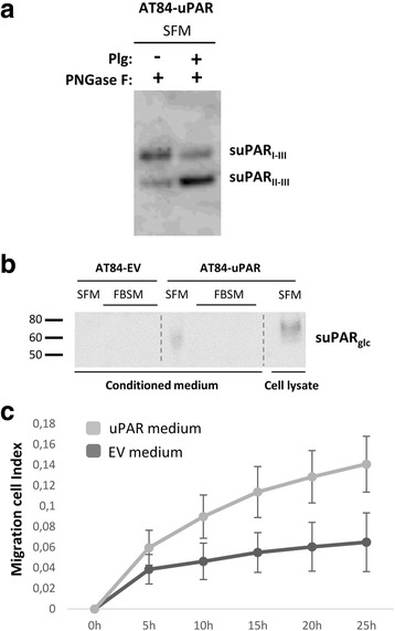
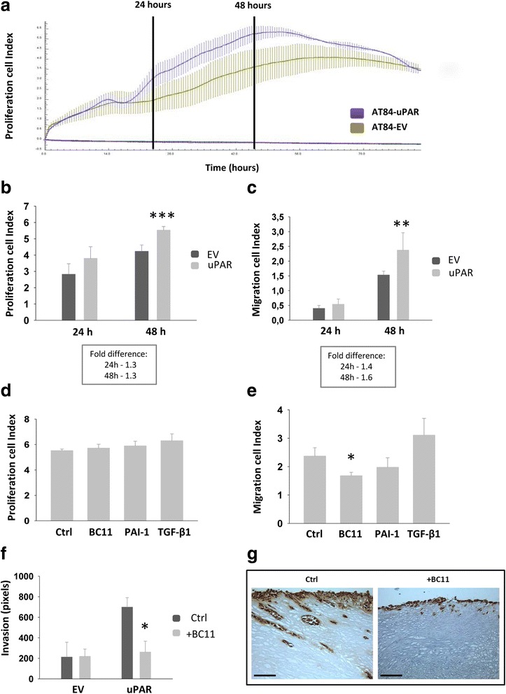
We found that when uPAR was overexpressed a proportion of the receptor was cleaved, thus the cells presented both full-length uPAR and uPAR (II-III). Cleavage was mainly performed by serine proteases and urokinase plasminogen activator (uPA) in particular. When the OSCC cells were stimulated with TGF-β1, the production of the uPA inhibitor PAI-1 was increased, resulting in a reduction of uPAR cleavage. By inhibiting cleavage of uPAR, cell migration was reduced, and by inhibiting uPA activity, invasion was reduced. We could also show that medium containing soluble uPAR (suPAR), and cleaved soluble uPAR (suPAR (II-III)), induced migration in OSCC cells with low endogenous levels of uPAR.

CONCLUSIONS

These results show that soluble factors in the tumour microenvironment, such as TGF-β1, PAI-1 and uPA, can influence the ratio of full length and uPAR (II-III) and thereby potentially effect cell migration and invasion. Resolving how uPAR cleavage is controlled is therefore vital for understanding how OSCC progresses and potentially provides new targets for therapy.

摘要

背景

尿激酶型纤溶酶原激活物(uPA)受体(uPAR)在人类口腔鳞状细胞癌(OSCC)的侵袭性肿瘤前沿上调,表明uPAR在肿瘤进展中发挥作用。我们之前在OSCC小鼠模型中观察到肿瘤-基质界面处uPAR表达升高,这与蛋白水解活性增加有关。肿瘤微环境调节uPAR的表达及其糖基化和裂解。全长和裂解的uPAR(uPAR(II-III))都参与细胞信号传导、增殖、迁移、干细胞动员和侵袭等高度调控的过程。本研究的目的是分析肿瘤相关因子及其对uPAR裂解的影响,以及对细胞增殖、迁移和侵袭的潜在影响。

方法

在小鼠OSCC细胞系AT84中稳定过表达小鼠uPAR。通过蛋白质印迹分析全长与裂解uPAR的比例,并通过添加不同的蛋白酶抑制剂和转化生长因子-β1(TGF-β1)评估其调控。使用实时细胞分析分析uPAR裂解在细胞增殖和迁移中的作用,并使用肌瘤侵袭模型评估侵袭。

结果

我们发现,当uPAR过表达时,一部分受体会被裂解,因此细胞同时呈现全长uPAR和uPAR(II-III)。裂解主要由丝氨酸蛋白酶特别是尿激酶型纤溶酶原激活物(uPA)进行。当用TGF-β1刺激OSCC细胞时,uPA抑制剂PAI-1的产生增加,导致uPAR裂解减少。通过抑制uPAR的裂解,细胞迁移减少,通过抑制uPA活性,侵袭减少。我们还可以表明,含有可溶性uPAR(suPAR)和裂解的可溶性uPAR(suPAR(II-III))的培养基可诱导内源性uPAR水平低的OSCC细胞迁移。

结论

这些结果表明,肿瘤微环境中的可溶性因子,如TGF-β1、PAI-1和uPA,可影响全长和uPAR(II-III)的比例,从而可能影响细胞迁移和侵袭。因此,解决uPAR裂解如何被控制对于理解OSCC的进展至关重要,并可能为治疗提供新的靶点。

https://cdn.ncbi.nlm.nih.gov/pmc/blobs/6a47/5438506/4802e6ab8400/12885_2017_3349_Fig4_HTML.jpg
https://cdn.ncbi.nlm.nih.gov/pmc/blobs/6a47/5438506/7033dfe13883/12885_2017_3349_Fig1_HTML.jpg
https://cdn.ncbi.nlm.nih.gov/pmc/blobs/6a47/5438506/e7f713a9fa64/12885_2017_3349_Fig2_HTML.jpg
https://cdn.ncbi.nlm.nih.gov/pmc/blobs/6a47/5438506/edcc7712f31c/12885_2017_3349_Fig3_HTML.jpg
https://cdn.ncbi.nlm.nih.gov/pmc/blobs/6a47/5438506/4802e6ab8400/12885_2017_3349_Fig4_HTML.jpg

相似文献

1
Cleavage of the urokinase receptor (uPAR) on oral cancer cells: regulation by transforming growth factor - β1 (TGF-β1) and potential effects on migration and invasion.口腔癌细胞上尿激酶受体(uPAR)的裂解:转化生长因子-β1(TGF-β1)的调节作用及其对迁移和侵袭的潜在影响
BMC Cancer. 2017 May 19;17(1):350. doi: 10.1186/s12885-017-3349-7.
2
Expression of urokinase-type plasminogen activator/urokinase-type plasminogen activator receptor and maspin in oral squamous cell carcinoma: Association with mode of invasion and clinicopathological factors.尿激酶型纤溶酶原激活物/尿激酶型纤溶酶原激活物受体和 maspin 在口腔鳞状细胞癌中的表达:与浸润方式和临床病理因素的关系。
Oncol Rep. 2011 Dec;26(6):1555-60. doi: 10.3892/or.2011.1419. Epub 2011 Aug 10.
3
TGF-β2 promotes RPE cell invasion into a collagen gel by mediating urokinase-type plasminogen activator (uPA) expression.TGF-β2 通过介导尿激酶型纤溶酶原激活物(uPA)的表达促进 RPE 细胞向胶原凝胶内浸润。
Exp Eye Res. 2013 Oct;115:13-21. doi: 10.1016/j.exer.2013.06.020. Epub 2013 Jun 28.
4
Tumour microenvironments induce expression of urokinase plasminogen activator receptor (uPAR) and concomitant activation of gelatinolytic enzymes.肿瘤微环境诱导尿激酶型纤溶酶原激活物受体(uPAR)的表达并伴随明胶酶的激活。
PLoS One. 2014 Aug 26;9(8):e105929. doi: 10.1371/journal.pone.0105929. eCollection 2014.
5
Targeting of urokinase plasminogen activator receptor in human pancreatic carcinoma cells inhibits c-Met- and insulin-like growth factor-I receptor-mediated migration and invasion and orthotopic tumor growth in mice.靶向人胰腺癌细胞中的尿激酶型纤溶酶原激活物受体可抑制c-Met和胰岛素样生长因子-I受体介导的迁移、侵袭以及小鼠原位肿瘤生长。
Cancer Res. 2005 Sep 1;65(17):7775-81. doi: 10.1158/0008-5472.CAN-05-0946.
6
Urinary-type plasminogen activator receptor/alpha 3 beta 1 integrin signaling, altered gene expression, and oral tumor progression.尿激酶型纤溶酶原激活物受体/α3β1 整联蛋白信号、基因表达改变与口腔肿瘤进展。
Mol Cancer Res. 2010 Feb;8(2):145-58. doi: 10.1158/1541-7786.MCR-09-0045. Epub 2010 Feb 9.
7
The urokinase-system in tumor tissue stroma of the breast and breast cancer cell invasion.乳腺肿瘤组织基质中的尿激酶系统与乳腺癌细胞侵袭
Int J Oncol. 2009 Jan;34(1):15-23.
8
Downregulation of urokinase plasminogen activator receptor expression inhibits Erk signalling with concomitant suppression of invasiveness due to loss of uPAR-beta1 integrin complex in colon cancer cells.尿激酶型纤溶酶原激活物受体表达的下调抑制了Erk信号传导,同时由于结肠癌细胞中uPAR-β1整合素复合物的缺失而抑制了侵袭性。
Br J Cancer. 2003 Jul 21;89(2):374-84. doi: 10.1038/sj.bjc.6601098.
9
Effects of transforming growth factor-β1 on plasminogen activation in stem cells from the apical papilla: role of activating receptor-like kinase 5/Smad2 and mitogen-activated protein kinase kinase (MEK)/extracellular signal-regulated kinase (ERK) signalling.转化生长因子-β1 对根尖乳头干细胞中纤溶酶原激活的影响:激活受体样激酶 5/Smad2 和丝裂原活化蛋白激酶激酶(MEK)/细胞外信号调节激酶(ERK)信号通路的作用。
Int Endod J. 2020 May;53(5):647-659. doi: 10.1111/iej.13266. Epub 2020 Feb 7.
10
Inhibition of epidermal growth factor-induced invasion by dexamethasone and AP-1 decoy in human squamous cell carcinoma cell lines.地塞米松和AP-1诱饵对人鳞状细胞癌细胞系中表皮生长因子诱导侵袭的抑制作用。
J Cell Physiol. 2002 Dec;193(3):340-8. doi: 10.1002/jcp.10181.

引用本文的文献

1
Changes in body composition in early breast cancer patients treated with aromatase inhibitors.接受芳香化酶抑制剂治疗的早期乳腺癌患者的身体成分变化。
J Endocrinol Invest. 2024 Dec;47(12):3119-3128. doi: 10.1007/s40618-024-02401-7. Epub 2024 Jun 10.
2
The Role of the Plasminogen/Plasmin System in Inflammation of the Oral Cavity.纤溶酶原/纤溶酶系统在口腔炎症中的作用。
Cells. 2023 Jan 30;12(3):445. doi: 10.3390/cells12030445.
3
Therapeutic and Metagenomic Potential of the Biomolecular Therapies against Periodontitis and the Oral Microbiome: Current Evidence and Future Perspectives.

本文引用的文献

1
Neoplastic extracellular matrix environment promotes cancer invasion in vitro.肿瘤细胞外基质环境在体外促进癌症侵袭。
Exp Cell Res. 2016 Jun 10;344(2):229-40. doi: 10.1016/j.yexcr.2016.04.003. Epub 2016 Apr 20.
2
Immunohistochemical Study of TGF-β1 in Oral Leukoplakia and Oral Squamous Cell Carcinoma: Correlations Between Clinicopathologic Factors and Overall Survival.转化生长因子-β1在口腔白斑及口腔鳞状细胞癌中的免疫组织化学研究:临床病理因素与总生存率的相关性
Appl Immunohistochem Mol Morphol. 2017 Oct;25(9):651-659. doi: 10.1097/PAI.0000000000000355.
3
Components of the Plasminogen-Plasmin System as Biologic Markers for Cancer.
生物分子疗法治疗牙周炎和口腔微生物组的治疗和宏基因组学潜力:当前证据和未来展望。
Int J Mol Sci. 2022 Nov 8;23(22):13708. doi: 10.3390/ijms232213708.
4
ALG3 Promotes Peritoneal Metastasis of Ovarian Cancer through Increasing Interaction of α1,3-mannosylated uPAR and ADAM8.ALG3 通过增加 α1,3-甘露糖化 uPAR 和 ADAM8 的相互作用促进卵巢癌细胞腹膜转移。
Cells. 2022 Oct 6;11(19):3141. doi: 10.3390/cells11193141.
5
An oxidative stress-related prognostic signature for indicating the immune status of oral squamous cell carcinoma and guiding clinical treatment.一种与氧化应激相关的预后特征,用于指示口腔鳞状细胞癌的免疫状态并指导临床治疗。
Front Genet. 2022 Sep 23;13:977902. doi: 10.3389/fgene.2022.977902. eCollection 2022.
6
Cancer-Associated Fibroblasts: Mechanisms of Tumor Progression and Novel Therapeutic Targets.癌症相关成纤维细胞:肿瘤进展机制与新型治疗靶点
Cancers (Basel). 2022 Feb 27;14(5):1231. doi: 10.3390/cancers14051231.
7
The Urokinase Plasminogen Activation System in Pancreatic Cancer: Prospective Diagnostic and Therapeutic Targets.尿激酶型纤溶酶原激活系统在胰腺癌中的作用:有前景的诊断和治疗靶点。
Biomolecules. 2022 Jan 18;12(2):152. doi: 10.3390/biom12020152.
8
Update on Novel Targeted Therapy for Pleural Organization and Fibrosis.胸膜组织和纤维化的新型靶向治疗进展。
Int J Mol Sci. 2022 Jan 29;23(3):1587. doi: 10.3390/ijms23031587.
9
uPAR: An Essential Factor for Tumor Development.尿激酶型纤溶酶原激活物受体:肿瘤发展的关键因素
J Cancer. 2021 Oct 17;12(23):7026-7040. doi: 10.7150/jca.62281. eCollection 2021.
10
TGF-β regulation of the uPA/uPAR axis modulates mesothelial-mesenchymal transition (MesoMT).TGF-β 对 uPA/uPAR 轴的调节可调节间皮-间充质转化(MesoMT)。
Sci Rep. 2021 Oct 27;11(1):21210. doi: 10.1038/s41598-021-99520-5.
纤溶酶原-纤溶酶系统的组成成分作为癌症的生物标志物
Adv Exp Med Biol. 2015;867:145-56. doi: 10.1007/978-94-017-7215-0_10.
4
Soluble Urokinase Receptor Is Released Selectively by Glioblastoma Cells That Express Epidermal Growth Factor Receptor Variant III and Promotes Tumor Cell Migration and Invasion.可溶性尿激酶受体由表达表皮生长因子受体变异体III的胶质母细胞瘤细胞选择性释放,并促进肿瘤细胞迁移和侵袭。
J Biol Chem. 2015 Jun 12;290(24):14798-809. doi: 10.1074/jbc.M115.637488. Epub 2015 Apr 2.
5
Tumour microenvironments induce expression of urokinase plasminogen activator receptor (uPAR) and concomitant activation of gelatinolytic enzymes.肿瘤微环境诱导尿激酶型纤溶酶原激活物受体(uPAR)的表达并伴随明胶酶的激活。
PLoS One. 2014 Aug 26;9(8):e105929. doi: 10.1371/journal.pone.0105929. eCollection 2014.
6
Urokinase plasminogen activator receptor (uPAR) and plasminogen activator inhibitor-1 (PAI-1) are potential predictive biomarkers in early stage oral squamous cell carcinomas (OSCC).尿激酶型纤溶酶原激活物受体(uPAR)和纤溶酶原激活物抑制剂-1(PAI-1)是早期口腔鳞状细胞癌(OSCC)潜在的预测生物标志物。
PLoS One. 2014 Jul 7;9(7):e101895. doi: 10.1371/journal.pone.0101895. eCollection 2014.
7
Expression of uPAR in tumor-associated stromal cells is associated with colorectal cancer patient prognosis: a TMA study.肿瘤相关基质细胞中尿激酶型纤溶酶原激活物受体(uPAR)的表达与结直肠癌患者预后相关:一项组织芯片研究
BMC Cancer. 2014 Apr 17;14:269. doi: 10.1186/1471-2407-14-269.
8
Insights into the role of components of the tumor microenvironment in oral carcinoma call for new therapeutic approaches.深入研究肿瘤微环境成分在口腔癌中的作用,需要寻求新的治疗方法。
Exp Cell Res. 2014 Jul 15;325(2):58-64. doi: 10.1016/j.yexcr.2013.12.029. Epub 2014 Jan 22.
9
Regulation of the anti-tumour immune response by cancer-associated fibroblasts.肿瘤相关成纤维细胞对肿瘤免疫应答的调控。
Semin Cancer Biol. 2014 Apr;25:69-77. doi: 10.1016/j.semcancer.2013.12.005. Epub 2014 Jan 7.
10
Identification of two distinct carcinoma-associated fibroblast subtypes with differential tumor-promoting abilities in oral squamous cell carcinoma.鉴定两种具有不同肿瘤促进能力的口腔鳞状细胞癌相关成纤维细胞亚型。
Cancer Res. 2013 Jul 1;73(13):3888-901. doi: 10.1158/0008-5472.CAN-12-4150. Epub 2013 Apr 18.