Sánchez-Catasús Carlos A, Sanabria-Diaz Gretel, Willemsen Antoon, Martinez-Montes Eduardo, Samper-Noa Juan, Aguila-Ruiz Angel, Boellaard Ronald, De Deyn Peter P, Dierckx Rudi A J O, Melie-Garcia Lester
Department of Nuclear Medicine and Molecular Imaging, University of Groningen, University Medical Center Groningen, The Netherlands; Department of Nuclear Medicine, Center for Neurological Restoration (CIREN), Havana, Cuba.
Laboratoire de Recherche en Neuroimagerie (LREN), Centre Hospitalier Universitaire Vaudois (CHUV), Lausanne, Switzerland; Neuroinformatics Department, Cuban Neuroscience Center, Havana, Cuba.
Neuroimage Clin. 2017 Apr 25;15:151-160. doi: 10.1016/j.nicl.2017.04.019. eCollection 2017.
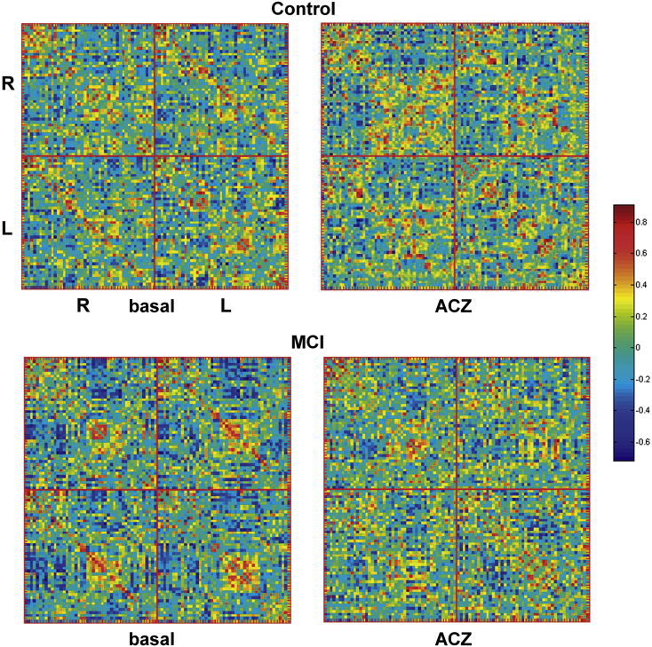
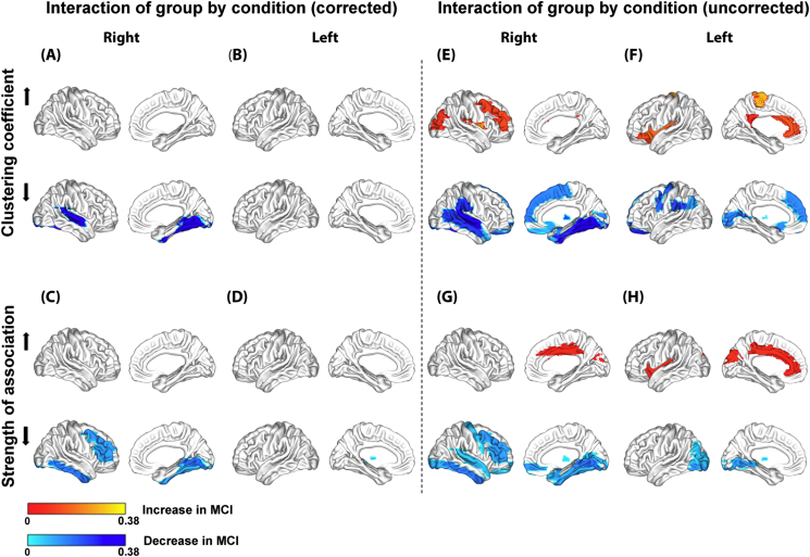
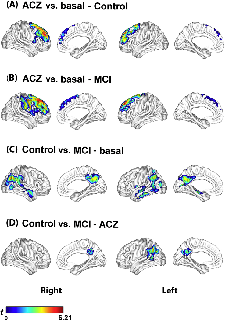
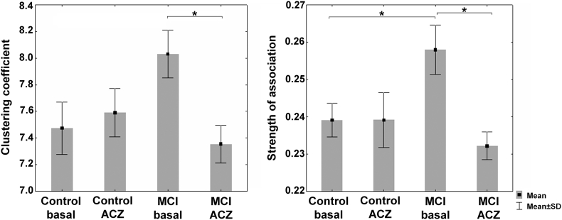
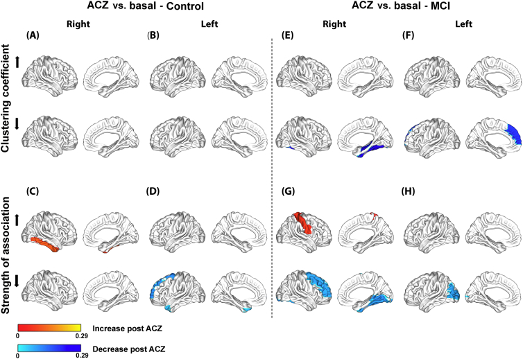
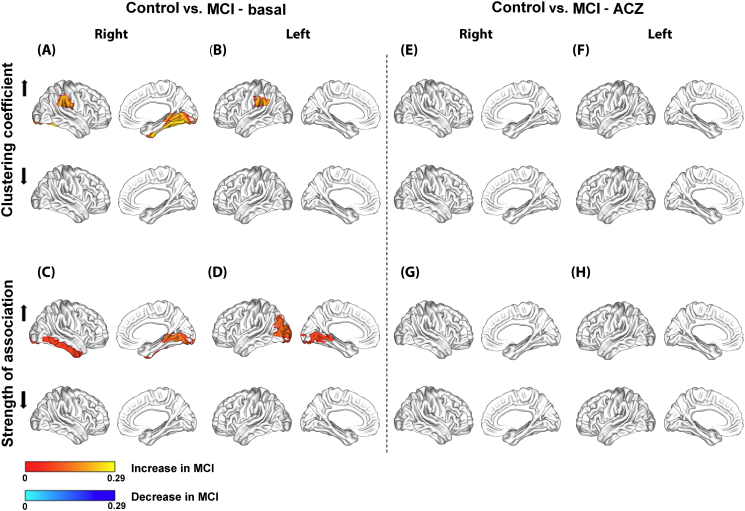
There is growing support that cerebrovascular reactivity (CVR) in response to a vasodilatory challenge, also defined as the cerebrovascular reserve, is reduced in Alzheimer's disease dementia. However, this is less clear in patients with mild cognitive impairment (MCI). The current standard analysis may not reflect subtle abnormalities in CVR. In this study, we aimed to investigate vasodilatory-induced changes in the topology of the cerebral blood flow correlation (CBF) network to study possible network-related CVR abnormalities in MCI. For this purpose, four CBF networks were constructed: two using CBF SPECT data at baseline and under the vasodilatory challenge of acetazolamide (ACZ), obtained from a group of 26 MCI patients; and two equivalent networks from a group of 26 matched cognitively normal controls. The mean strength of association () and clustering coefficient () were used to evaluate ACZ-induced changes on the topology of CBF networks. We found that cognitively normal adults and MCI patients show different patterns of and changes. The observed differences included the medial prefrontal cortices and inferior parietal lobe, which represent areas involved in MCI's cognitive dysfunction. In contrast, no substantial differences were detected by standard CVR analysis. These results suggest that graph theoretical analysis of ACZ-induced changes in the topology of the CBF networks allows the identification of subtle network-related CVR alterations in MCI, which couldn't be detected by the standard approach.
越来越多的证据表明,在阿尔茨海默病痴呆症患者中,对血管舒张刺激的脑血管反应性(CVR),也被定义为脑血管储备,会降低。然而,在轻度认知障碍(MCI)患者中,情况尚不清楚。目前的标准分析可能无法反映CVR的细微异常。在本研究中,我们旨在研究血管舒张诱导的脑血流相关性(CBF)网络拓扑结构变化,以探讨MCI中可能存在的与网络相关的CVR异常。为此,构建了四个CBF网络:两个使用来自26名MCI患者组在基线和乙酰唑胺(ACZ)血管舒张刺激下的CBF单光子发射计算机断层扫描(SPECT)数据;另外两个等效网络来自26名匹配的认知正常对照组。使用平均关联强度()和聚类系数()来评估ACZ诱导的CBF网络拓扑结构变化。我们发现,认知正常成年人和MCI患者表现出不同的 和 变化模式。观察到的差异包括内侧前额叶皮质和顶下叶,这些区域与MCI的认知功能障碍有关。相比之下,标准CVR分析未检测到实质性差异。这些结果表明,对ACZ诱导的CBF网络拓扑结构变化进行图论分析,可以识别出MCI中与网络相关的细微CVR改变,而这是标准方法无法检测到的。