• 文献检索
  • 文档翻译
  • 深度研究
  • 学术资讯
  • Suppr Zotero 插件Zotero 插件
  • 邀请有礼
  • 套餐&价格
  • 历史记录
应用&插件
Suppr Zotero 插件Zotero 插件浏览器插件Mac 客户端Windows 客户端微信小程序
定价
高级版会员购买积分包购买API积分包
服务
文献检索文档翻译深度研究API 文档MCP 服务
关于我们
关于 Suppr公司介绍联系我们用户协议隐私条款
关注我们

Suppr 超能文献

核心技术专利:CN118964589B侵权必究
粤ICP备2023148730 号-1Suppr @ 2026

文献检索

告别复杂PubMed语法,用中文像聊天一样搜索,搜遍4000万医学文献。AI智能推荐,让科研检索更轻松。

立即免费搜索

文件翻译

保留排版,准确专业,支持PDF/Word/PPT等文件格式,支持 12+语言互译。

免费翻译文档

深度研究

AI帮你快速写综述,25分钟生成高质量综述,智能提取关键信息,辅助科研写作。

立即免费体验

胰腺神经内分泌肿瘤中基于影像学和病理学维度的比较。

Comparison of imaging-based and pathological dimensions in pancreatic neuroendocrine tumors.

作者信息

Paiella Salvatore, Impellizzeri Harmony, Zanolin Elisabetta, Marchegiani Giovanni, Miotto Marco, Malpaga Anna, De Robertis Riccardo, D'Onofrio Mirko, Rusev Borislav, Capelli Paola, Cingarlini Sara, Butturini Giovanni, Davì Maria Vittoria, Amodio Antonio, Bassi Claudio, Scarpa Aldo, Salvia Roberto, Landoni Luca

机构信息

Salvatore Paiella, Harmony Impellizzeri, Giovanni Marchegiani, Marco Miotto, Anna Malpaga, Claudio Bassi, Roberto Salvia, Luca Landoni, General and Pancreatic Surgery Department, Pancreas Institute, University and Hospital Trust of Verona, 37134 Verona, Italy.

出版信息

World J Gastroenterol. 2017 May 7;23(17):3092-3098. doi: 10.3748/wjg.v23.i17.3092.

DOI:10.3748/wjg.v23.i17.3092
PMID:28533666
原文链接:https://pmc.ncbi.nlm.nih.gov/articles/PMC5423046/
Abstract

AIM

To establish the ability of magnetic resonance (MR) and computer tomography (CT) to predict pathologic dimensions of pancreatic neuroendocrine tumors (PanNET) in a caseload of a tertiary referral center.

METHODS

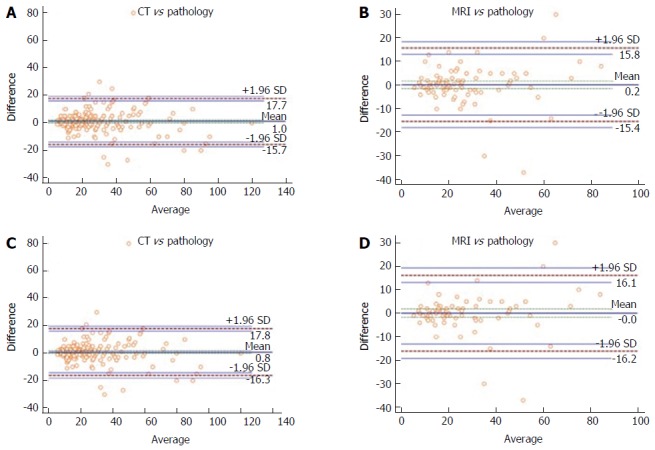
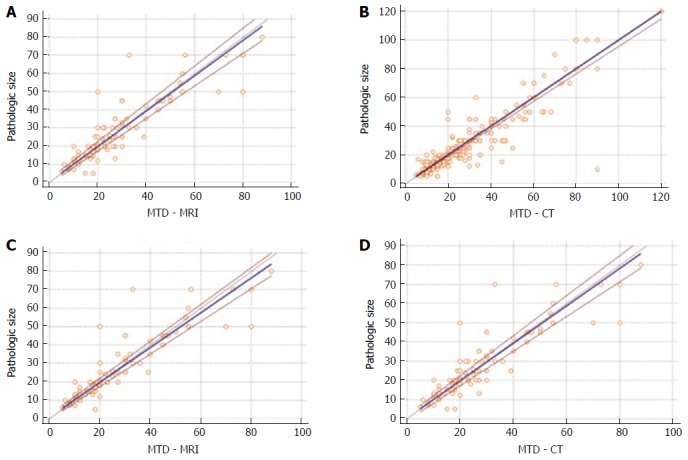
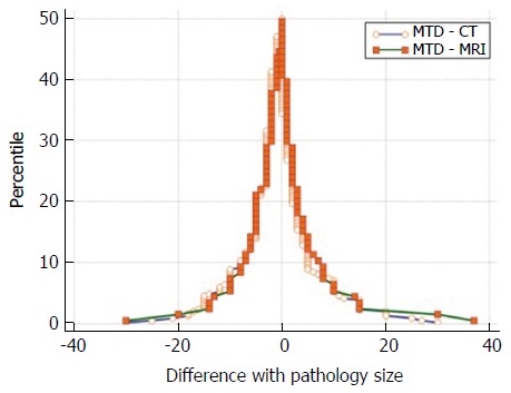
Patients submitted to surgery for PanNET at the Surgical Unit of the Pancreas Institute with at least 1 preoperative imaging examination (MR or CT scan) from January 2005 to December 2015 were included and data retrospectively collected. Exclusion criteria were: multifocal lesions, genetic syndromes, microadenomas or mixed tumors, metastatic disease and neoadjuvant therapy. Bland-Altman (BA) and Mountain-Plot (MP) statistics were used to compare size measured by each modality with the pathology size. Passing-Bablok (PB) regression analysis was used to check the agreement between MR and CT.

RESULTS

Our study population consisted of 292 patients. Seventy-nine (27.1%) were functioning PanNET. The mean biases were 0.17 ± 7.99 mm, 1 ± 8.51 mm and 0.23 ± 9 mm, 1.2 ± 9.8 mm for MR and CT, considering the overall population and the subgroup of non-functioning- PanNET, respectively. Limits of agreement (LOA) included the vast majority of observations, indicating a good agreement between imaging and pathology. The MP further confirmed this finding and showed that the two methods are unbiased with respect to each other. Considering ≤ 2 cm non-functioning-PanNET, no statistical significance was found in the size estimation rate of MR and CT ( = 0.433). PBR analysis did not reveal significant differences between MR, CT and pathology.

CONCLUSION

MR and CT scan are accurate and interchangeable imaging techniques in predicting pathologic dimensions of PanNET.

摘要

目的

在一家三级转诊中心的病例中,确定磁共振成像(MR)和计算机断层扫描(CT)预测胰腺神经内分泌肿瘤(PanNET)病理大小的能力。

方法

纳入2005年1月至2015年12月期间在胰腺研究所外科接受PanNET手术且术前至少进行过1次影像学检查(MR或CT扫描)的患者,并回顾性收集数据。排除标准为:多灶性病变、遗传综合征、微腺瘤或混合瘤、转移性疾病和新辅助治疗。采用布兰德-奥特曼(BA)统计和山形图(MP)统计来比较每种检查方式测得的大小与病理大小。采用帕辛-巴布洛(PB)回归分析来检验MR和CT之间的一致性。

结果

我们的研究队列包括292例患者。79例(27.1%)为功能性PanNET。对于总体人群和无功能性PanNET亚组,MR的平均偏差分别为0.17±7.99毫米、1±8.51毫米,CT的平均偏差分别为0.23±9毫米、1.2±9.8毫米。一致性界限(LOA)包括了绝大多数观察值,表明影像学与病理之间具有良好的一致性。MP进一步证实了这一发现,并表明这两种方法相互之间无偏差。对于直径≤2厘米的无功能性PanNET,MR和CT在大小估计率方面未发现统计学差异(P = 0.433)。PB回归分析未显示MR、CT与病理之间存在显著差异。

结论

MR和CT扫描在预测PanNET病理大小方面是准确且可相互替代的影像学技术。

https://cdn.ncbi.nlm.nih.gov/pmc/blobs/3955/5423046/18d72a883c8e/WJG-23-3092-g003.jpg
https://cdn.ncbi.nlm.nih.gov/pmc/blobs/3955/5423046/4c81e0930eee/WJG-23-3092-g001.jpg
https://cdn.ncbi.nlm.nih.gov/pmc/blobs/3955/5423046/d0853cd1455a/WJG-23-3092-g002.jpg
https://cdn.ncbi.nlm.nih.gov/pmc/blobs/3955/5423046/18d72a883c8e/WJG-23-3092-g003.jpg
https://cdn.ncbi.nlm.nih.gov/pmc/blobs/3955/5423046/4c81e0930eee/WJG-23-3092-g001.jpg
https://cdn.ncbi.nlm.nih.gov/pmc/blobs/3955/5423046/d0853cd1455a/WJG-23-3092-g002.jpg
https://cdn.ncbi.nlm.nih.gov/pmc/blobs/3955/5423046/18d72a883c8e/WJG-23-3092-g003.jpg

相似文献

1
Comparison of imaging-based and pathological dimensions in pancreatic neuroendocrine tumors.胰腺神经内分泌肿瘤中基于影像学和病理学维度的比较。
World J Gastroenterol. 2017 May 7;23(17):3092-3098. doi: 10.3748/wjg.v23.i17.3092.
2
Pancreatic neuroendocrine tumor: Correlations between MRI features, tumor biology, and clinical outcome after surgery.胰腺神经内分泌肿瘤:MRI 特征、肿瘤生物学与手术治疗后临床结局的相关性。
J Magn Reson Imaging. 2018 Feb;47(2):425-432. doi: 10.1002/jmri.25756. Epub 2017 May 8.
3
Feasibility of Radio-Guided Surgery with ⁶⁸Gallium-DOTATATE in Patients with Gastro-Entero-Pancreatic Neuroendocrine Tumors.⁶⁸镓-奥曲肽在胃肠胰神经内分泌肿瘤患者中进行放射性引导手术的可行性
Ann Surg Oncol. 2015 Dec;22 Suppl 3(Suppl 3):S676-82. doi: 10.1245/s10434-015-4857-9. Epub 2015 Sep 8.
4
Identification of primary tumors in patients presenting with metastatic gastroenteropancreatic neuroendocrine tumors.转移性胃肠胰神经内分泌肿瘤患者中原发性肿瘤的鉴定
Surgery. 2017 Jan;161(1):272-279. doi: 10.1016/j.surg.2016.05.055. Epub 2016 Nov 15.
5
99mTc-tektrotyd SPECT/MRI in the diagnosis of a gastroenteropancreatic neuroendocrine tumor.99mTc-替曲膦单光子发射计算机断层扫描/磁共振成像在胃肠胰神经内分泌肿瘤诊断中的应用
Clin Nucl Med. 2012 Apr;37(4):e84-5. doi: 10.1097/RLU.0b013e3182478a7e.
6
A rare case with synchronous gastric gastrointestinal stromal tumor, pancreatic neuroendocrine tumor, and uterine leiomyoma.一例同时患有胃胃肠道间质瘤、胰腺神经内分泌肿瘤和子宫平滑肌瘤的罕见病例。
World J Surg Oncol. 2016 Nov 15;14(1):287. doi: 10.1186/s12957-016-1051-x.
7
Pancreatic neuroendocrine tumor in a patient with a TSC1 variant: case report and review of the literature.一名携带TSC1变异体患者的胰腺神经内分泌肿瘤:病例报告及文献综述
Fam Cancer. 2018 Apr;17(2):275-280. doi: 10.1007/s10689-017-0029-3.
8
Clinical Usefulness of Somatostatin Receptor Scintigraphy in Japanese Patients with Gastroenteropancreatic Neuroendocrine Tumors.生长抑素受体闪烁扫描术在日本胃肠胰神经内分泌肿瘤患者中的临床应用价值
Digestion. 2017;96(1):13-20. doi: 10.1159/000470838. Epub 2017 Jun 14.
9
Is the combination of MR and CT findings useful in determining the tumor grade of pancreatic neuroendocrine tumors?磁共振成像(MR)和计算机断层扫描(CT)检查结果的组合对于确定胰腺神经内分泌肿瘤的肿瘤分级是否有用?
Jpn J Radiol. 2017 May;35(5):242-253. doi: 10.1007/s11604-017-0627-x. Epub 2017 Mar 3.
10
Dual tracer functional imaging of gastroenteropancreatic neuroendocrine tumors using 68Ga-DOTA-NOC PET-CT and 18F-FDG PET-CT: competitive or complimentary?使用 68Ga-DOTA-NOC PET-CT 和 18F-FDG PET-CT 对胃肠胰神经内分泌肿瘤进行双示踪剂功能成像:竞争还是互补?
Clin Nucl Med. 2014 Jan;39(1):e27-34. doi: 10.1097/RLU.0b013e31827a216b.

引用本文的文献

1
Preoperative Prediction of Lymph Node Metastases in Nonfunctional Pancreatic Neuroendocrine Tumors Using a Combined CT Radiomics-Clinical Model.使用 CT 影像组学-临床联合模型预测无功能性胰腺神经内分泌肿瘤的淋巴结转移。
Ann Surg Oncol. 2024 Nov;31(12):8136-8145. doi: 10.1245/s10434-024-16064-4. Epub 2024 Aug 23.
2
PANCREATODUODENECTOMY IN PATIENT WITH VON HIPPEL-LINDAU DISEASE: A LITERATURE REVIEW.《von Hippel-Lindau 病患者行胰十二指肠切除术:文献复习》
Arq Bras Cir Dig. 2022 Nov 25;35:e1697. doi: 10.1590/0102-672020220002e1697. eCollection 2022.
3
NETest: serial liquid biopsies in gastroenteropancreatic NET surveillance.

本文引用的文献

1
ENETS Consensus Guidelines Update for the Management of Patients with Functional Pancreatic Neuroendocrine Tumors and Non-Functional Pancreatic Neuroendocrine Tumors.《欧洲神经内分泌肿瘤学会功能性胰腺神经内分泌肿瘤和非功能性胰腺神经内分泌肿瘤患者管理共识指南更新》
Neuroendocrinology. 2016;103(2):153-71. doi: 10.1159/000443171. Epub 2016 Jan 5.
2
Pancreatic Neuroendocrine Neoplasms: Clinical Value of Diffusion-Weighted Imaging.胰腺神经内分泌肿瘤:扩散加权成像的临床价值
Neuroendocrinology. 2016;103(6):758-70. doi: 10.1159/000442984. Epub 2015 Dec 9.
3
Is the 2-cm size cutoff relevant for small nonfunctioning pancreatic neuroendocrine tumors: A French multicenter study.
NET检测:胃肠胰神经内分泌肿瘤监测中的系列液体活检
Endocr Connect. 2022 Sep 7;11(10). doi: 10.1530/EC-22-0146. Print 2022 Oct 1.
4
[ELF4 promotes proliferation and inhibits apoptosis of human insulinoma cells by activating Akt signaling].[ELF4通过激活Akt信号通路促进人胰岛素瘤细胞增殖并抑制其凋亡]
Nan Fang Yi Ke Da Xue Xue Bao. 2021 Aug 31;41(9):1329-1333. doi: 10.12122/j.issn.1673-4254.2021.09.06.
5
Pancreatic neuroendocrine tumours: spectrum of imaging findings.胰腺神经内分泌肿瘤:影像学表现谱
Gland Surg. 2020 Dec;9(6):2215-2224. doi: 10.21037/gs-20-537.
6
Reliability and Agreement of Radiological and Pathological Tumor Size in Patients with Multiple Endocrine Neoplasia Type 1-Related Pancreatic Neuroendocrine Tumors: Results from a Population-Based Cohort.多内分泌腺瘤病 1 型相关胰腺神经内分泌肿瘤患者的影像学和病理学肿瘤大小的可靠性和一致性:基于人群的队列研究结果。
Neuroendocrinology. 2021;111(8):705-717. doi: 10.1159/000510514. Epub 2020 Jul 28.
2厘米大小的临界值对小型无功能胰腺神经内分泌肿瘤是否具有相关性:一项法国多中心研究。
Surgery. 2016 Mar;159(3):901-7. doi: 10.1016/j.surg.2015.10.003. Epub 2015 Nov 14.
4
Cystic pancreatic neuroendocrine tumors: outcomes of preoperative endosonography-guided fine needle aspiration, and recurrence during long-term follow-up.胰腺囊性神经内分泌肿瘤:术前超声内镜引导下细针抽吸的结果,以及长期随访期间的复发情况。
Endoscopy. 2015 Jul;47(7):617-25. doi: 10.1055/s-0034-1391712. Epub 2015 Mar 12.
5
Pancreatic neuroendocrine tumors: accurate grading with Ki-67 index on fine-needle aspiration specimens using the WHO 2010/ENETS criteria.胰腺神经内分泌肿瘤:使用 WHO 2010/ENETS 标准,通过细针穿刺标本中的 Ki-67 指数进行准确分级。
Cancer Cytopathol. 2014 Oct;122(10):770-8. doi: 10.1002/cncy.21457. Epub 2014 Jul 9.
6
Observational study of natural history of small sporadic nonfunctioning pancreatic neuroendocrine tumors.散发性无功能性小胰腺神经内分泌肿瘤自然史的观察性研究。
J Clin Endocrinol Metab. 2013 Dec;98(12):4784-9. doi: 10.1210/jc.2013-2604. Epub 2013 Sep 20.
7
Small, nonfunctioning, asymptomatic pancreatic neuroendocrine tumors (PNETs): role for nonoperative management.体积小、无功能、无症状的胰腺神经内分泌肿瘤(PNET):非手术治疗的作用。
Surgery. 2012 Dec;152(6):965-74. doi: 10.1016/j.surg.2012.08.038. Epub 2012 Oct 24.
8
Ki-67 grading of nonfunctioning pancreatic neuroendocrine tumors on histologic samples obtained by EUS-guided fine-needle tissue acquisition: a prospective study.EUS 引导下细针组织获取的组织样本中 Ki-67 分级在非功能性胰腺神经内分泌肿瘤中的应用:一项前瞻性研究。
Gastrointest Endosc. 2012 Sep;76(3):570-7. doi: 10.1016/j.gie.2012.04.477.
9
ENETS Consensus Guidelines for the management of patients with digestive neuroendocrine neoplasms of the digestive system: well-differentiated pancreatic non-functioning tumors.ENETS消化系统消化神经内分泌肿瘤患者管理共识指南:高分化胰腺无功能肿瘤
Neuroendocrinology. 2012;95(2):120-34. doi: 10.1159/000335587. Epub 2012 Feb 15.
10
Tumor size correlates with malignancy in nonfunctioning pancreatic endocrine tumor.肿瘤大小与无功能性胰腺内分泌肿瘤的恶性程度相关。
Surgery. 2011 Jul;150(1):75-82. doi: 10.1016/j.surg.2011.02.022.