• 文献检索
  • 文档翻译
  • 深度研究
  • 学术资讯
  • Suppr Zotero 插件Zotero 插件
  • 邀请有礼
  • 套餐&价格
  • 历史记录
应用&插件
Suppr Zotero 插件Zotero 插件浏览器插件Mac 客户端Windows 客户端微信小程序
定价
高级版会员购买积分包购买API积分包
服务
文献检索文档翻译深度研究API 文档MCP 服务
关于我们
关于 Suppr公司介绍联系我们用户协议隐私条款
关注我们

Suppr 超能文献

核心技术专利:CN118964589B侵权必究
粤ICP备2023148730 号-1Suppr @ 2026

文献检索

告别复杂PubMed语法,用中文像聊天一样搜索,搜遍4000万医学文献。AI智能推荐,让科研检索更轻松。

立即免费搜索

文件翻译

保留排版,准确专业,支持PDF/Word/PPT等文件格式,支持 12+语言互译。

免费翻译文档

深度研究

AI帮你快速写综述,25分钟生成高质量综述,智能提取关键信息,辅助科研写作。

立即免费体验

眼前节光学相干断层扫描与伪装成慢性前葡萄膜炎的残留植物性眼内异物

Anterior segment optical coherence tomography and retained vegetal intraocular foreign body masquerading as chronic anterior uveitis.

作者信息

Mahmoud Anis, Messaoud Riadh, Abid Fatma, Ksiaa Imen, Bouzayene Melek, Khairallah Moncef

机构信息

Department of Ophthalmology, Taher Sfar University Hospital, Faculty of Medicine, University of Monastir, Monastir, Tunisia.

Department of Ophthalmology, Fattouma Bourguiba University Hospital, Faculty of Medicine, University of Monastir, 5019, Monastir, Tunisia.

出版信息

J Ophthalmic Inflamm Infect. 2017 Dec;7(1):13. doi: 10.1186/s12348-017-0130-7. Epub 2017 May 23.

DOI:10.1186/s12348-017-0130-7
PMID:28536985
原文链接:https://pmc.ncbi.nlm.nih.gov/articles/PMC5442073/
Abstract

BACKGROUND

The purpose of this single case report was to report the use of anterior segment optical coherence tomography for the diagnosis and management of a retained vegetal intraocular foreign body.

RESULTS

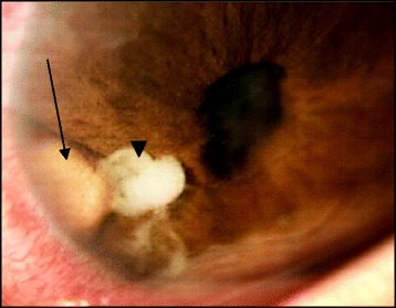
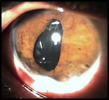
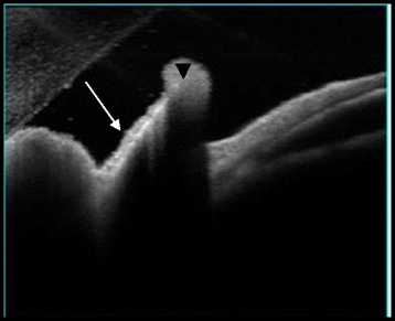
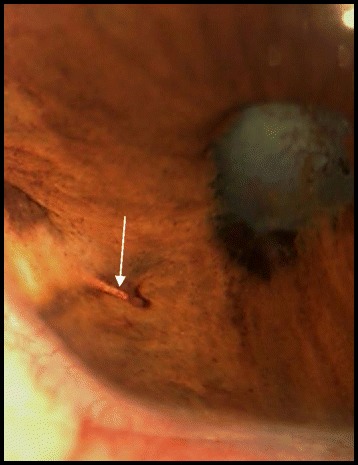
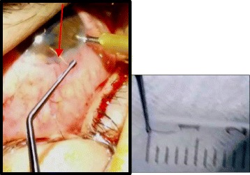
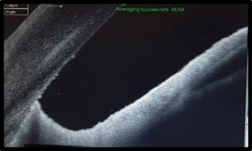
A 23-year-old otherwise healthy male presented with a progressive vision loss in the right eye (RE). He reported a mild ocular trauma with a tree leaf 1 year ago followed by recurrent episodes of redness and pain in the RE that partially resolved after a self-medication with topical steroids. Visual acuity of the RE was limited to light perception. Slit-lamp examination of the RE showed an iris granuloma with overlying exudate and associated anterior chamber inflammatory reaction. Film X-rays, contact B-scan ultrasonography, and CT scan showed no abnormalities. Anterior segment optical coherence tomography revealed an enclaved iris foreign body. The foreign body was removed after a short course of local antibio-corticosteroid therapy. This was followed 2 months later by cataract surgery with intraocular lens implantation, with subsequent improvement of visual acuity to 20/40.

CONCLUSIONS

A missed intraocular foreign body can lead to sight-threatening complications. Anterior segment optical coherence tomography may be useful for detecting non-clinically evident intraocular foreign body involving the anterior segment masquerading as chronic anterior uveitis.

摘要

背景

本单病例报告旨在阐述眼前节光学相干断层扫描技术在诊断和处理滞留植物性眼内异物中的应用。

结果

一名23岁身体健康的男性因右眼视力进行性下降前来就诊。他自述1年前眼部曾被树叶轻微划伤,之后右眼反复出现眼红、疼痛,自行使用局部类固醇药物治疗后症状部分缓解。右眼视力仅存光感。裂隙灯检查发现右眼有虹膜肉芽肿伴上方渗出物及相关的前房炎症反应。X线平片、接触式B超及CT扫描均未发现异常。眼前节光学相干断层扫描显示有一枚包埋于虹膜的异物。经短期局部抗生素-皮质类固醇治疗后取出异物。2个月后行白内障摘除联合人工晶状体植入术,术后视力提高至20/40。

结论

眼内异物漏诊可导致威胁视力的并发症。眼前节光学相干断层扫描技术对于检测伪装成慢性前葡萄膜炎的非临床显性眼前节眼内异物可能有用。

https://cdn.ncbi.nlm.nih.gov/pmc/blobs/97c7/5442073/c907e744c917/12348_2017_130_Fig6_HTML.jpg
https://cdn.ncbi.nlm.nih.gov/pmc/blobs/97c7/5442073/4ce2bf82a5a3/12348_2017_130_Fig1_HTML.jpg
https://cdn.ncbi.nlm.nih.gov/pmc/blobs/97c7/5442073/29a75be11044/12348_2017_130_Fig2_HTML.jpg
https://cdn.ncbi.nlm.nih.gov/pmc/blobs/97c7/5442073/39496dfbbbdb/12348_2017_130_Fig3_HTML.jpg
https://cdn.ncbi.nlm.nih.gov/pmc/blobs/97c7/5442073/e2bbb171d80a/12348_2017_130_Fig4_HTML.jpg
https://cdn.ncbi.nlm.nih.gov/pmc/blobs/97c7/5442073/8a7d602de8fe/12348_2017_130_Fig5_HTML.jpg
https://cdn.ncbi.nlm.nih.gov/pmc/blobs/97c7/5442073/c907e744c917/12348_2017_130_Fig6_HTML.jpg

相似文献

1
Anterior segment optical coherence tomography and retained vegetal intraocular foreign body masquerading as chronic anterior uveitis.眼前节光学相干断层扫描与伪装成慢性前葡萄膜炎的残留植物性眼内异物
J Ophthalmic Inflamm Infect. 2017 Dec;7(1):13. doi: 10.1186/s12348-017-0130-7. Epub 2017 May 23.
2
Anterior segment intraocular metallic foreign body causing chronic hypopyon uveitis.眼前节眼内金属异物致慢性前房积脓性葡萄膜炎。
J Ophthalmic Inflamm Infect. 2011 Jun;1(2):85-7. doi: 10.1007/s12348-010-0011-9. Epub 2010 Nov 17.
3
Role of B-scan ultrasonography in the localization of intraocular foreign bodies in the anterior segment: a report of three cases.B超在眼前段眼内异物定位中的作用:三例报告
BMC Ophthalmol. 2015 Aug 14;15:102. doi: 10.1186/s12886-015-0076-1.
4
Missed intraocular foreign body masquerading as intraocular inflammation: two cases.
Int Ophthalmol. 2010 Dec;30(6):713-6. doi: 10.1007/s10792-010-9350-7. Epub 2010 Feb 27.
5
Lens siderosis resulting from a tiny missed intralenticular foreign body.因一个微小的晶状体内异物未被发现而导致的晶状体铁锈症。
J Chin Med Assoc. 2009 Jan;72(1):42-4. doi: 10.1016/S1726-4901(09)70019-X.
6
A missed diagnosis of multiple intraocular foreign bodies for 21 years.21年来多发性眼内异物漏诊
Cont Lens Anterior Eye. 2017 Dec;40(6):432-435. doi: 10.1016/j.clae.2017.08.002. Epub 2017 Aug 24.
7
[Foreign Body in the Anterior Chamber 10 Months after Uneventful Cataract Surgery].
Nippon Ganka Gakkai Zasshi. 2016 Apr;120(4):310-5.
8
Occult anterior-chamber metallic fragment post-phacoemulsification masquerading as chronic recalcitrant postoperative inflammation.白内障超声乳化术后隐匿性前房金属碎片伪装成慢性顽固性术后炎症。
Am J Ophthalmol. 2005 Mar;139(3):541-2. doi: 10.1016/j.ajo.2004.08.031.
9
Spontaneous Scleral Perforation of an Anterior Chamber Intraocular Lens.前房人工晶状体自发性巩膜穿孔
Case Rep Ophthalmol. 2016 May 9;7(1):249-52. doi: 10.1159/000445880. eCollection 2016 Jan-Apr.
10
Case Report: Evaluating Intraocular Foreign Bodies After Corneal Perforation Using Swept Source Anterior Segment Optical Coherence Tomography.病例报告:使用扫频源眼前节光学相干断层扫描评估角膜穿孔后的眼内异物
Optom Vis Sci. 2020 Feb;97(2):101-103. doi: 10.1097/OPX.0000000000001475.

引用本文的文献

1
Clinical Characteristics, Outcomes, and Complications Associated With Delayed Diagnosis of Intraocular Foreign Body.眼内异物延迟诊断的临床特征、结局及并发症
J Vitreoretin Dis. 2025 May 29:24741264251339090. doi: 10.1177/24741264251339090.
2
Intraocular foreign body presenting as recalcitrant hypopyon anterior uveitis.表现为顽固性前房积脓性前葡萄膜炎的眼内异物。
Taiwan J Ophthalmol. 2021 Apr 17;13(1):114-116. doi: 10.4103/tjo.tjo_1_21. eCollection 2023 Jan-Mar.
3
Role of AS-OCT in Managing Corneal Disorders.眼前节光学相干断层扫描在角膜疾病管理中的作用。

本文引用的文献

1
The role of anterior segment optical coherence tomography in the management of an intra-corneal foreign body.眼前节光学相干断层扫描在角膜异物处理中的作用
Springerplus. 2016 Sep 13;5(1):1559. doi: 10.1186/s40064-016-3242-x. eCollection 2016.
2
Intraocular foreign bodies: A review.眼内异物:综述
Surv Ophthalmol. 2016 Sep-Oct;61(5):582-96. doi: 10.1016/j.survophthal.2016.03.005. Epub 2016 Mar 17.
3
Identification and localization of multiple intrastromal foreign bodies with anterior segment optical coherence tomography and ocular Pentacam.
Diagnostics (Basel). 2022 Apr 7;12(4):918. doi: 10.3390/diagnostics12040918.
4
Unnoticed metallic foreign body in the camerular angle inducing chronic uveitis.房角内未被注意到的金属异物导致慢性葡萄膜炎。
Arq Bras Oftalmol. 2021 May-Jun;84(3):267-270. doi: 10.5935/0004-2749.20210038.
5
Unusually located metallic foreign body in posterior chamber of eyeball: role of multimodal ocular imaging in its diagnosis and management.眼球后房内位置异常的金属异物:多模态眼部成像在其诊断和处理中的作用
BMJ Case Rep. 2020 Oct 27;13(10):e239345. doi: 10.1136/bcr-2020-239345.
使用眼前节光学相干断层扫描和眼前节分析仪对多个基质内异物进行识别和定位。
Int Ophthalmol. 2014 Apr;34(2):355-8. doi: 10.1007/s10792-013-9800-0. Epub 2013 Jun 6.
4
Anterior segment intraocular metallic foreign body causing chronic hypopyon uveitis.眼前节眼内金属异物致慢性前房积脓性葡萄膜炎。
J Ophthalmic Inflamm Infect. 2011 Jun;1(2):85-7. doi: 10.1007/s12348-010-0011-9. Epub 2010 Nov 17.
5
Missed intraocular foreign body masquerading as intraocular inflammation: two cases.
Int Ophthalmol. 2010 Dec;30(6):713-6. doi: 10.1007/s10792-010-9350-7. Epub 2010 Feb 27.
6
Optical coherence tomography in the detection of an intraocular foreign body.
Acta Ophthalmol. 2009 Aug;87(5):582-3. doi: 10.1111/j.1755-3768.2008.01242.x. Epub 2008 Oct 17.