• 文献检索
  • 文档翻译
  • 深度研究
  • 学术资讯
  • Suppr Zotero 插件Zotero 插件
  • 邀请有礼
  • 套餐&价格
  • 历史记录
应用&插件
Suppr Zotero 插件Zotero 插件浏览器插件Mac 客户端Windows 客户端微信小程序
定价
高级版会员购买积分包购买API积分包
服务
文献检索文档翻译深度研究API 文档MCP 服务
关于我们
关于 Suppr公司介绍联系我们用户协议隐私条款
关注我们

Suppr 超能文献

核心技术专利:CN118964589B侵权必究
粤ICP备2023148730 号-1Suppr @ 2026

文献检索

告别复杂PubMed语法,用中文像聊天一样搜索,搜遍4000万医学文献。AI智能推荐,让科研检索更轻松。

立即免费搜索

文件翻译

保留排版,准确专业,支持PDF/Word/PPT等文件格式,支持 12+语言互译。

免费翻译文档

深度研究

AI帮你快速写综述,25分钟生成高质量综述,智能提取关键信息,辅助科研写作。

立即免费体验

Presurgical role of MRI tractography in a case of extensive cervicothoracic spinal ependymoma.

作者信息

Granata Francesca, Racchiusa Sergio, Mormina Enricomaria, Barresi Valeria, Garufi Giada, Grasso Giovanni, Salpietro Francesco Maria, Longo Marcello, Alafaci Concetta

机构信息

Neuroradiology Unit, Department of Biomedical Sciences and Morphological and Functional Imaging, University of Messina, Messina, Italy.

Section of Pathological Anatomy, Department of Human Pathology, University of Messina, Messina, Italy.

出版信息

Surg Neurol Int. 2017 Apr 26;8:56. doi: 10.4103/sni.sni_33_17. eCollection 2017.

DOI:10.4103/sni.sni_33_17
PMID:28540122
原文链接:https://pmc.ncbi.nlm.nih.gov/articles/PMC5421221/
Abstract

BACKGROUND

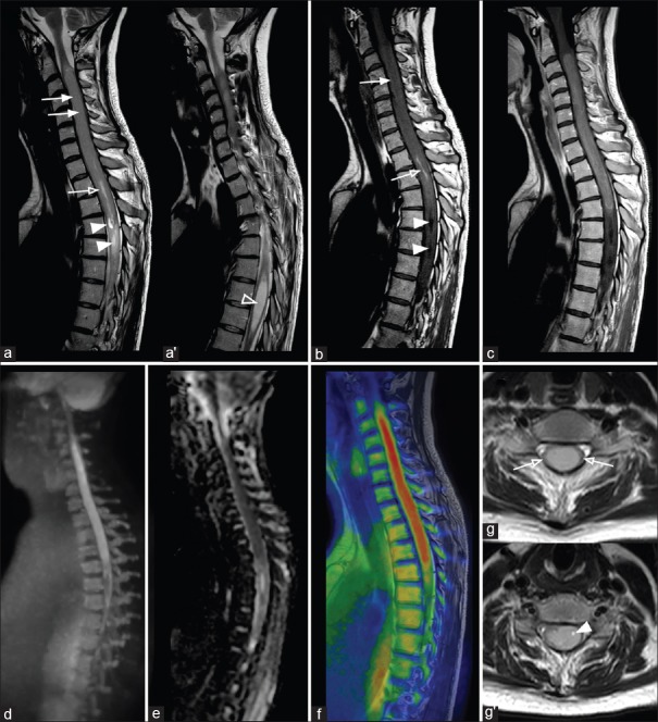
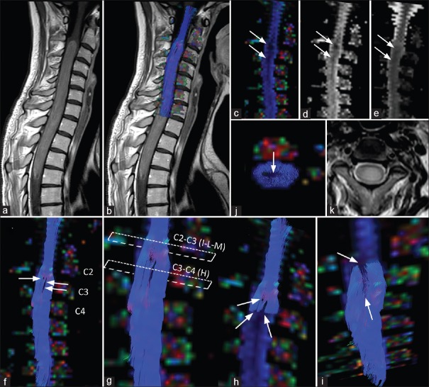
Intramedullary spinal ependymoma is a tumor, hardly characterizable with conventional magnetic resonance (MR) imaging only. MR diffusion tensor imaging (DTI) with three-dimensional fiber-tracking reconstructions allows the evaluation of the relationship between neoplasm and white matter fiber tracts, being a powerful tool in presurgical planning. We present DTI findings in a case of a young female with an extensive cervicothoracic spinal ependymoma.

CASE DESCRIPTION

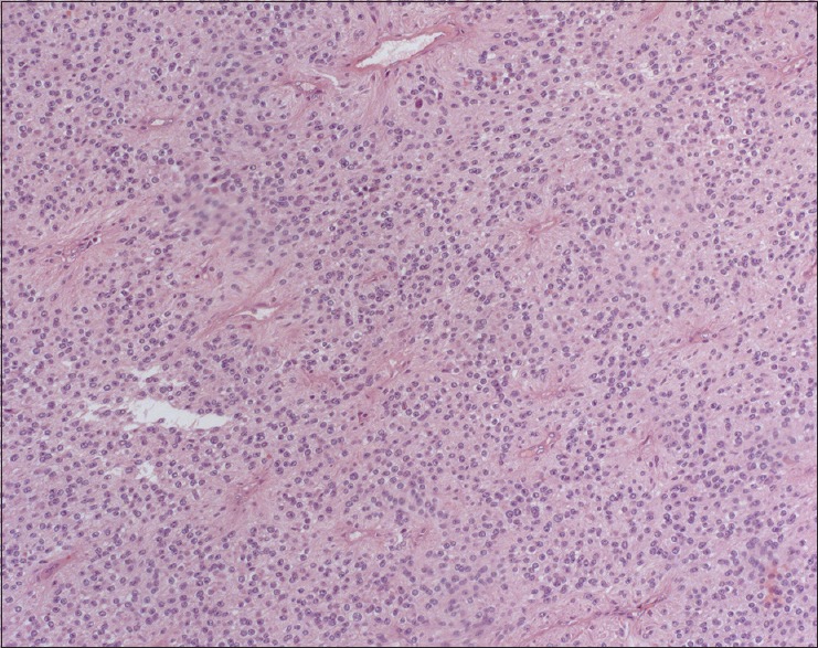
The patient complained of a 2-month history of acute urinary retention, weakness and numbness on the lower limbs and the upper left limb. She underwent MR imaging that showed an extensive cervicothoracic spinal mass, difficult to characterize with conventional MR sequences. DTI showed peripherally displacement of fibers, without involvement of the spinal cord, findings consistent with an ependymoma. The patient underwent surgery with a complete resection "" of the lesion, which showed clear cleavage planes, as detected by DTI. Histopathological findings confirmed the diagnosis of ependymoma.

CONCLUSIONS

DTI is a useful tool in presurgical planning, helping in differentiating not infiltrating neoplasms, such as spinal ependymomas, from other infiltrative and more aggressive neoplasms, which are considered not resectable.

摘要
https://cdn.ncbi.nlm.nih.gov/pmc/blobs/8dd6/5421221/92d16e349263/SNI-8-56-g003.jpg
https://cdn.ncbi.nlm.nih.gov/pmc/blobs/8dd6/5421221/08adf51c076d/SNI-8-56-g001.jpg
https://cdn.ncbi.nlm.nih.gov/pmc/blobs/8dd6/5421221/85863861a901/SNI-8-56-g002.jpg
https://cdn.ncbi.nlm.nih.gov/pmc/blobs/8dd6/5421221/92d16e349263/SNI-8-56-g003.jpg
https://cdn.ncbi.nlm.nih.gov/pmc/blobs/8dd6/5421221/08adf51c076d/SNI-8-56-g001.jpg
https://cdn.ncbi.nlm.nih.gov/pmc/blobs/8dd6/5421221/85863861a901/SNI-8-56-g002.jpg
https://cdn.ncbi.nlm.nih.gov/pmc/blobs/8dd6/5421221/92d16e349263/SNI-8-56-g003.jpg

相似文献

1
Presurgical role of MRI tractography in a case of extensive cervicothoracic spinal ependymoma.
Surg Neurol Int. 2017 Apr 26;8:56. doi: 10.4103/sni.sni_33_17. eCollection 2017.
2
Combined Use of Diffusion Tractography and Advanced Intraoperative Imaging for Resection of Cervical Intramedullary Spinal Cord Neoplasms: A Case Series and Technical Note.弥散张量成像与高级术中成像在颈髓内肿瘤切除术中的联合应用:病例系列及技术探讨。
Oper Neurosurg (Hagerstown). 2019 Nov 1;17(5):525-530. doi: 10.1093/ons/opz039.
3
Microsurgery resection of giant cervicothoracic spinal ependymoma: Two-dimensional operative video.巨大型颈胸段脊髓室管膜瘤的显微手术切除:二维手术视频
Surg Neurol Int. 2024 Jul 12;15:237. doi: 10.25259/SNI_317_2024. eCollection 2024.
4
Spinal diffusion tensor tractography for differentiation of intramedullary tumor-suspected lesions.用于鉴别髓内疑似肿瘤病变的脊髓扩散张量纤维束成像
Eur J Radiol. 2016 Dec;85(12):2275-2280. doi: 10.1016/j.ejrad.2016.10.018. Epub 2016 Oct 17.
5
Conus Medullaris Teratoma with Utilization of Fiber Tractography: Case Report.圆锥马尾畸胎瘤与纤维束成像技术的应用:病例报告
J Neurol Surg Rep. 2015 Jul;76(1):e183-7. doi: 10.1055/s-0035-1555134. Epub 2015 Jun 12.
6
'Serpent in the spine': a case of giant spinal ependymoma of cervicothoracic spine.“脊柱中的蛇”:一例颈胸段巨大脊髓室管膜瘤
BMJ Case Rep. 2012 Jun 27;2012:5890. doi: 10.1136/bcr-02-2012-5890.
7
[Comparative analysis between multi-segments intramedullary spinal cord congenital tumors and benign ependymomas].[多节段脊髓先天性肿瘤与良性室管膜瘤的对比分析]
Beijing Da Xue Xue Bao Yi Xue Ban. 2010 Apr 18;42(2):183-7.
8
Diffusion tensor tractography of residual fibers in traumatic spinal cord injury: a pilot study.创伤性脊髓损伤后残留纤维的弥散张量纤维束追踪:一项初步研究。
J Neuroradiol. 2013 Jul;40(3):181-6. doi: 10.1016/j.neurad.2012.08.008. Epub 2013 Feb 18.
9
Efficacy of diffusion tensor anisotropy indices and tractography in assessing the extent of severity of spinal cord injury: an in vitro analytical study in calf spinal cords.弥散张量各向异性指数和轨迹在评估脊髓损伤严重程度中的作用:牛脊髓的体外分析研究。
Spine J. 2012 Dec;12(12):1147-53. doi: 10.1016/j.spinee.2012.10.032. Epub 2012 Dec 16.
10
Surgical experience with resection en bloc of intramedullary astrocytomas and ependymomas in the cervical and cervicothoracic region.颈段及颈胸段髓内星形细胞瘤和室管膜瘤整块切除的手术经验。
J Clin Neurosci. 2004 Aug;11(6):623-8. doi: 10.1016/j.jocn.2003.08.011.

引用本文的文献

1
The use of diffusion tensor imaging in spinal pathology: a comprehensive literature review.弥散张量成像在脊柱病变中的应用:全面文献综述。
Eur Spine J. 2024 Sep;33(9):3303-3314. doi: 10.1007/s00586-024-08231-8. Epub 2024 Jul 16.
2
Full cervical cord tractography: A new method for clinical use.全颈髓纤维束成像:一种临床应用的新方法。
Front Neuroanat. 2022 Sep 27;16:993464. doi: 10.3389/fnana.2022.993464. eCollection 2022.
3
Surgical approaches to intramedullary spinal cord astrocytomas in the age of genomics.基因组学时代脊髓髓内星形细胞瘤的手术治疗方法

本文引用的文献

1
Spinal diffusion tensor tractography for differentiation of intramedullary tumor-suspected lesions.用于鉴别髓内疑似肿瘤病变的脊髓扩散张量纤维束成像
Eur J Radiol. 2016 Dec;85(12):2275-2280. doi: 10.1016/j.ejrad.2016.10.018. Epub 2016 Oct 17.
2
Diffusion tensor imaging to guide surgical planning in intramedullary spinal cord tumors in children.扩散张量成像用于指导儿童脊髓髓内肿瘤的手术规划。
Neuroradiology. 2014 Feb;56(2):169-74. doi: 10.1007/s00234-013-1316-9. Epub 2014 Jan 7.
3
Advanced MR diffusion tensor imaging and perfusion weighted imaging of intramedullary tumors and tumor like lesions in the cervicomedullary junction region and the cervical spinal cord.
Front Oncol. 2022 Sep 6;12:982089. doi: 10.3389/fonc.2022.982089. eCollection 2022.
4
Sodium Fluorescein for Spinal Intradural Tumors.用于脊髓硬膜内肿瘤的荧光素钠
Front Oncol. 2021 Jan 28;10:618579. doi: 10.3389/fonc.2020.618579. eCollection 2020.
颈髓交界区及颈段脊髓内肿瘤样病变的高级磁共振弥散张量成像及灌注加权成像。
J Neurooncol. 2014 Feb;116(3):559-66. doi: 10.1007/s11060-013-1323-z. Epub 2013 Dec 30.
4
Deterministic diffusion fiber tracking improved by quantitative anisotropy.通过定量各向异性改进的确定性扩散纤维追踪
PLoS One. 2013 Nov 15;8(11):e80713. doi: 10.1371/journal.pone.0080713. eCollection 2013.
5
Clinical applications of diffusion tensor imaging.弥散张量成像的临床应用。
World Neurosurg. 2014 Jul-Aug;82(1-2):96-109. doi: 10.1016/j.wneu.2013.07.083. Epub 2013 Aug 3.
6
[Spinal cord diffusion tensor tractography as a diagnostic tool in difficult cases of spinal cord intramedullary tumours].
Neurol Neurochir Pol. 2013 Jan-Feb;47(1):74-9. doi: 10.5114/ninp.2013.32938.
7
Diffusion tensor imaging tractography in patients with intramedullary tumors: comparison with intraoperative findings and value for prediction of tumor resectability.脊髓内肿瘤患者的弥散张量成像纤维束追踪:与术中发现的比较及对肿瘤可切除性预测的价值。
J Neurosurg Spine. 2010 Sep;13(3):371-80. doi: 10.3171/2010.3.SPINE09399.
8
Diffusion-weighted imaging, diffusion-tensor imaging, and fiber tractography of the spinal cord.脊髓的扩散加权成像、扩散张量成像和纤维束成像。
Magn Reson Imaging Clin N Am. 2009 May;17(2):225-44. doi: 10.1016/j.mric.2009.02.004.
9
Clinical applications of diffusion tensor tractography of the spinal cord.脊髓弥散张量纤维束成像的临床应用
Neuroradiology. 2008 Jan;50(1):25-9. doi: 10.1007/s00234-007-0309-y. Epub 2007 Oct 2.
10
Diffusion tensor magnetic resonance imaging and fiber tracking in spinal cord lesions: current and future indications.脊髓病变的扩散张量磁共振成像与纤维追踪:当前及未来的应用指征
Neuroimaging Clin N Am. 2007 Feb;17(1):137-47. doi: 10.1016/j.nic.2006.11.005.