Institute of Clinical Medicine, School of Medicine, National Yang-Ming University, Taipei, Taiwan.
Department of Orthopaedics & Traumatology, Taipei Veterans General Hospital, Taiwan.
Stem Cells Transl Med. 2017 Jun;6(6):1504-1514. doi: 10.1002/sctm.15-0394.
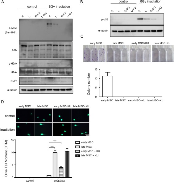
Cell therapies using human mesenchymal stem cells (MSCs) have received much attention in the past decade. In pursuit of the therapeutic potential of MSCs, cell expansion is required to generate a great number of cells with desired phenotype and functionality. Long-term expansion in vitro, however, can lead to altered functions. To explore the changes in DNA damage responses (DDR) in MSCs expanded, DDR pathways following irradiation were characterized in early- and late-passage bone marrow MSCs. Seventy-two hours after irradiation, the percentage of sub-G1 cells in early-passage MSCs did not change significantly. Reduced TUNEL staining was observed in early-passage MSCs compared to late-passage MSCs 4 h after irradiation. Comet assay also revealed that early-passage MSCs were more resistant to irradiation or DNA damages induced by genotoxic agents than late-passage MSCs. ATM phosphorylation and γ-H2AX and phospho-p53 increased in early-passage MSCs while decreased in late-passage MSCs. Through inhibition by KU55933, DDR pathway in early-passage MSCs was shown to be ATM-dependent. Higher levels of poly (ADP-ribose) polymerase-1 (PARP-1) and PAR synthesis were observed in early-passage MSCs than in late-passage MSCs. Knockdown of PARP-1 in early-passage MSCs resulted in sensitization to irradiation-induced apoptosis. Overexpression of PARP-1 in late passage MSCs could render irradiation resistance. Lower activity of DDR in late-passage MSCs was associated with rapid proteasomal degradation of PARP-1. In conclusion, early-passage MSCs are more irradiation-resistant and have increased DDR activity involving PARP-1, ATM and their downstream signals. Stem Cells Translational Medicine 2017;6:1504-1514.
在过去的十年中,使用人间质干细胞(MSCs)的细胞疗法受到了广泛关注。为了追求 MSC 的治疗潜力,需要进行细胞扩增以产生具有所需表型和功能的大量细胞。然而,长期体外扩增会导致功能改变。为了探讨扩增的 MSC 中 DNA 损伤反应(DDR)的变化,研究人员在早期和晚期传代骨髓 MSC 中表征了照射后的 DDR 途径。照射后 72 小时,早期传代 MSC 中的亚 G1 细胞百分比没有明显变化。与晚期传代 MSC 相比,照射后 4 小时,早期传代 MSC 中的 TUNEL 染色减少。彗星试验还表明,与晚期传代 MSC 相比,早期传代 MSC 对照射或遗传毒性药物诱导的 DNA 损伤更具抵抗力。早期传代 MSC 中的 ATM 磷酸化和 γ-H2AX 和磷酸化 p53 增加,而晚期传代 MSC 中的则减少。通过 KU55933 抑制,早期传代 MSC 中的 DDR 途径被证明是 ATM 依赖性的。早期传代 MSC 中的多聚(ADP-核糖)聚合酶-1(PARP-1)和 PAR 合成水平高于晚期传代 MSC。早期传代 MSC 中 PARP-1 的敲低导致对辐射诱导的细胞凋亡敏感。晚期传代 MSC 中 PARP-1 的过表达可使照射具有抗性。晚期传代 MSC 中 DDR 活性较低与 PARP-1 的快速蛋白酶体降解有关。总之,早期传代 MSC 对辐射的抵抗力更强,并且 DDR 活性增加,涉及 PARP-1、ATM 及其下游信号。干细胞转化医学 2017;6:1504-1514。