Kern Kyle C, Wright Clinton B, Bergfield Kaitlin L, Fitzhugh Megan C, Chen Kewei, Moeller James R, Nabizadeh Nooshin, Elkind Mitchell S V, Sacco Ralph L, Stern Yaakov, DeCarli Charles S, Alexander Gene E
Department of Neurology, Evelyn F. McKnight Brain Institute, University of Miami Miller School of MedicineMiami, FL, USA.
Neuroscience and Physiological Sciences Graduate Interdisciplinary Programs, University of ArizonaTucson, AZ, USA.
Front Aging Neurosci. 2017 May 15;9:132. doi: 10.3389/fnagi.2017.00132. eCollection 2017.
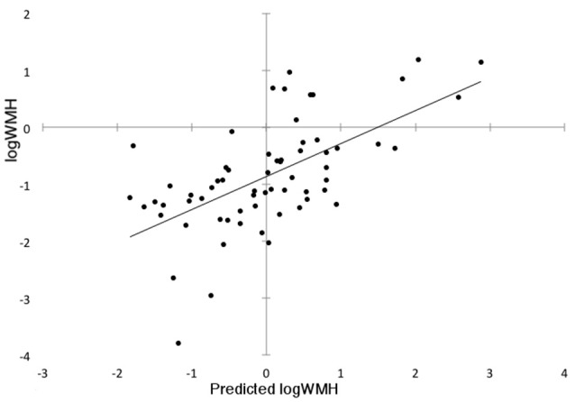
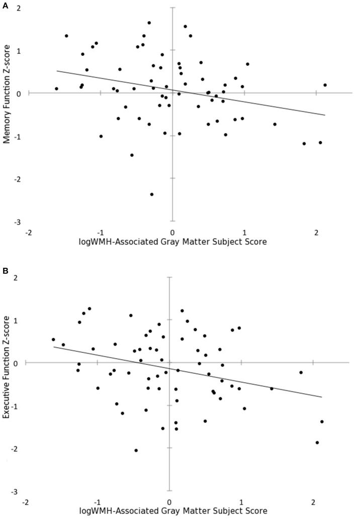
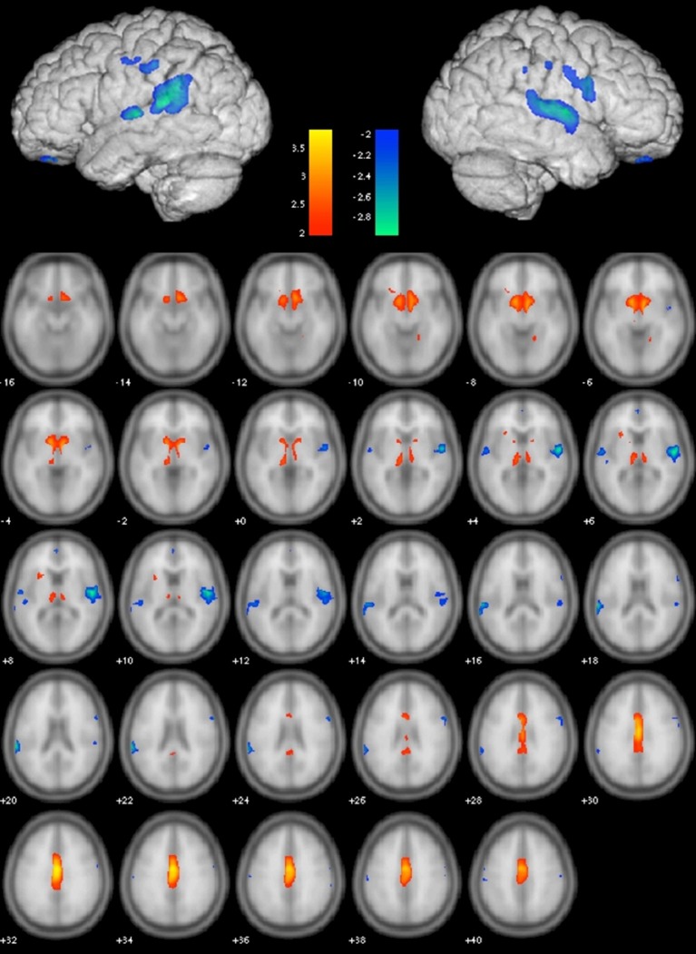
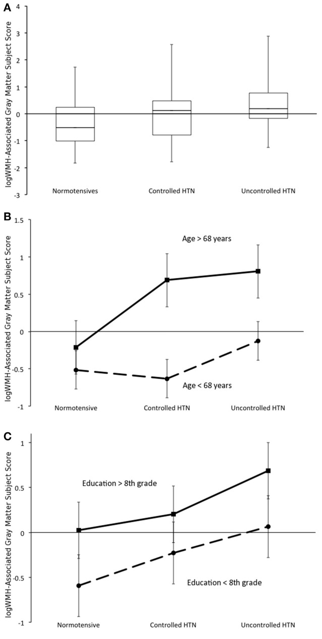
Cerebral small-vessel damage manifests as white matter hyperintensities and cerebral atrophy on brain MRI and is associated with aging, cognitive decline and dementia. We sought to examine the interrelationship of these imaging biomarkers and the influence of hypertension in older individuals. We used a multivariate spatial covariance neuroimaging technique to localize the effects of white matter lesion load on regional gray matter volume and assessed the role of blood pressure control, age and education on this relationship. Using a case-control design matching for age, gender, and educational attainment we selected 64 participants with normal blood pressure, controlled hypertension or uncontrolled hypertension from the Northern Manhattan Study cohort. We applied gray matter voxel-based morphometry with the scaled subprofile model to (1) identify regional covariance patterns of gray matter volume differences associated with white matter lesion load, (2) compare this relationship across blood pressure groups, and (3) relate it to cognitive performance. In this group of participants aged 60-86 years, we identified a pattern of reduced gray matter volume associated with white matter lesion load in bilateral temporal-parietal regions with relative preservation of volume in the basal forebrain, thalami and cingulate cortex. This pattern was expressed most in the uncontrolled hypertension group and least in the normotensives, but was also more evident in older and more educated individuals. Expression of this pattern was associated with worse performance in executive function and memory. In summary, white matter lesions from small-vessel disease are associated with a regional pattern of gray matter atrophy that is mitigated by blood pressure control, exacerbated by aging, and associated with cognitive performance.
脑小血管损伤在脑部磁共振成像(MRI)上表现为白质高信号和脑萎缩,与衰老、认知衰退及痴呆相关。我们试图研究这些影像学生物标志物之间的相互关系以及高血压对老年人的影响。我们使用多变量空间协方差神经成像技术来定位白质病变负荷对区域灰质体积的影响,并评估血压控制、年龄和教育程度在这种关系中的作用。采用病例对照设计,匹配年龄、性别和教育程度,我们从北曼哈顿研究队列中选取了64名血压正常、高血压得到控制或未得到控制的参与者。我们应用基于体素的灰质形态测量法和缩放子轮廓模型来(1)识别与白质病变负荷相关的灰质体积差异的区域协方差模式,(2)比较不同血压组之间的这种关系,以及(3)将其与认知表现相关联。在这组年龄在60 - 86岁的参与者中,我们发现双侧颞顶叶区域存在与白质病变负荷相关的灰质体积减少模式,而基底前脑、丘脑和扣带回皮质的体积相对保留。这种模式在未控制高血压组中表现最为明显,在血压正常组中表现最不明显,但在年龄较大和受教育程度较高的个体中也更明显。这种模式的表现与执行功能和记忆方面较差的表现相关。总之,小血管疾病导致的白质病变与一种灰质萎缩的区域模式相关,这种模式可通过血压控制得到缓解,随衰老而加剧,并与认知表现相关。