Verma Sudeep, Chidambaratanu Shanthi, Vijaylakshmi Raja, Srinivasan Latha, Suresh Indrani
Department of Paediatric Cardiac Sciences, Krishna Institute of MedicalSciences (KIMS), Secunderabad, Telangana, India.
Fetal Cardiology and Fetal Medicine Unit, Mediscan Systems, Mylapore, Chennai, Tamil Nadu, India.
Ann Pediatr Cardiol. 2017 May-Aug;10(2):215-217. doi: 10.4103/apc.APC_10_17.
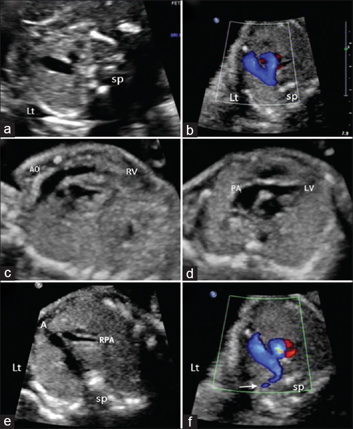
Transposition of great arteries (TGA) is more commonly associated with D-malposition of great arteries where anterior aorta produces characteristic "I" sign in the three-vessel view (3VV) in fetal heart imaging. We describe two cases with TGA and L-malposition of aorta where 3VV imaging showed an apparently normal arrangement of vessels while outflow tract imaging proved vital in diagnosing transposition anatomy. Apparently, normal 3VV in the presence of disproportionate vessel caliber and inability to produce normal outflow images should raise the suspicion. Attempts should be made to produce views to show great arteries originating from respective ventricles to rule out ventriculoarterial discordance and to complete segmental analysis.
大动脉转位(TGA)更常与大动脉右位心相关,在胎儿心脏成像的三血管切面(3VV)中,前方的主动脉会产生特征性的“ I”征。我们描述了两例伴有TGA和主动脉左位心的病例,其中3VV成像显示血管排列明显正常,而流出道成像对诊断转位解剖结构至关重要。显然,在血管口径不成比例且无法产生正常流出图像的情况下,3VV看似正常应引起怀疑。应尝试获得显示大动脉分别起源于各自心室的图像,以排除心室动脉不一致,并完成节段分析。