Wang Jia-Song, Xie Hua-Tao, Zhang Ming-Chang
Department of Ophthalmology, Union Hospital, Tongji Medical College, Huazhong University of Science and Technology, Wuhan 430022, China.
J Ophthalmol. 2017;2017:6761714. doi: 10.1155/2017/6761714. Epub 2017 May 8.
To ex vivo expand oral mucosal epithelium cells (OMECs) on acellular porcine corneal stroma (APCS) without using feeder cells and serum and to compare the morphologic and phenotypic characteristics of cultured oral cells on APCS to those of cells on deluded human amniotic membrane (HAM).
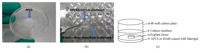
SD rat oral mucosal biopsies were cultured on APCS and HAM. Reverse-transcription polymerase chain reaction (RT-PCR) and immunohistochemistry were used to analyze the characterization of stem cells and epithelial differentiation of the outgrowth products.
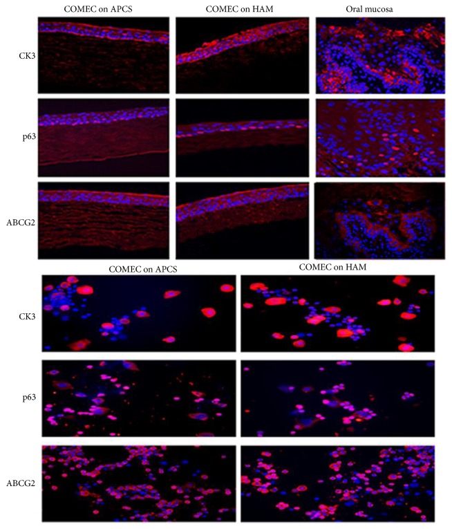
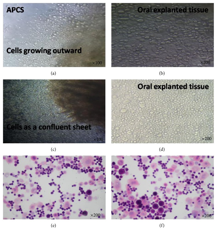
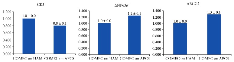
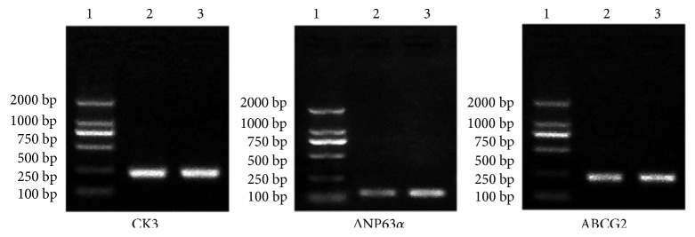
Stratified and optimal transplantable OMECs were obtained after being cultured three to four weeks. Both RT-PCR and immunohistochemistry showed that cultured OMECs expressed markers of epithelial differentiation cytokeratin K3 and epithelial stem cell markers of p63 and ABCG2.
OMECs can be successfully cultured on APCS without using xenobiotic feeder cells and serum. Characterization showed that these sheets retain the morphologic and phenotypic characteristics of OMECs within differentiated cells and stem cells. The optimal transplantable sheets can prove to be particularly beneficial to both bilateral limbal stem cell deficiency and deep corneal lesions.
在不使用饲养细胞和血清的情况下,在脱细胞猪角膜基质(APCS)上对口腔黏膜上皮细胞(OMECs)进行体外扩增,并比较在APCS上培养的口腔细胞与在去上皮人羊膜(HAM)上培养的细胞的形态和表型特征。
将SD大鼠口腔黏膜活检组织在APCS和HAM上培养。采用逆转录聚合酶链反应(RT-PCR)和免疫组织化学方法分析生长产物的干细胞特征和上皮分化情况。
培养三到四周后获得了分层且可用于移植的最佳OMECs。RT-PCR和免疫组织化学均显示,培养的OMECs表达上皮分化标志物细胞角蛋白K3以及上皮干细胞标志物p63和ABCG2。
无需使用异种饲养细胞和血清即可在APCS上成功培养OMECs。特征分析表明,这些细胞片保留了分化细胞和干细胞中OMECs的形态和表型特征。最佳可移植细胞片对双侧角膜缘干细胞缺乏症和深层角膜病变均可能特别有益。