Horiguchi Keishi, Tosaka Masahiko, Higuchi Tetsuya, Arisaka Yukiko, Sugawara Kenichi, Hirato Junko, Yokoo Hideaki, Tsushima Yoshito, Yoshimoto Yuhei
Department of Neurosurgery, Gunma University Graduate School of Medicine, 3-39-22 Showa-machi, Maebashi, Gunma, 371-8511, Japan.
Department of Diagnostic Radiology and Nuclear Medicine, Gunma University Graduate School of Medicine, Maebashi, Gunma, Japan.
EJNMMI Res. 2017 Dec;7(1):50. doi: 10.1186/s13550-017-0298-8. Epub 2017 May 31.
We investigated the relationship between metabolic activity and histological features of gliomas using fluorine-18α-methyltyrosine (F-FAMT) positron emission tomography (PET) compared with fluorine-18 fluorodeoxyglucose (F-FDG) PET in 38 consecutive glioma patients. The tumor to normal brain ratios (T/N ratios) were calculated, and the relationships between T/N ratio and World Health Organization tumor grade or MIB-1 labeling index were evaluated. The diagnostic values of T/N ratios were assessed using receiver operating characteristic (ROC) curve analyses to differentiate between high-grade gliomas (HGGs) and low-grade gliomas (LGGs).
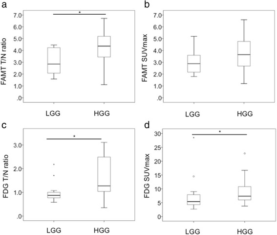
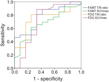
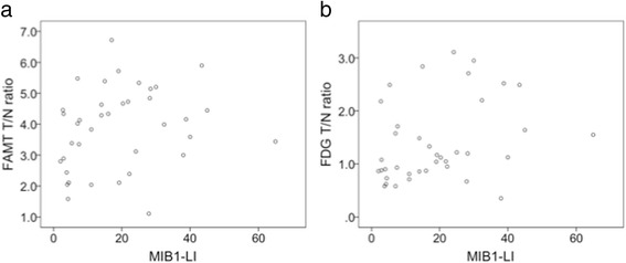
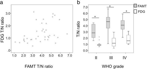
Median T/N ratio of F-FAMT PET was 2.85, 4.65, and 4.09 for grade II, III, and IV gliomas, respectively, with significant differences between HGGs and LGGs (p = 0.006). Both T/N ratio (p = 0.016) and maximum standardized uptake value (p = 0.033) of F-FDG PET showed significant differences between HGGs and LGGs. ROC analysis yielded an optimal cut-off of 3.37 for the T/N ratio of F-FAMT PET to differentiate between HGGs and LGGs (sensitivity 81%, specificity 67%, accuracy 76%, area under the ROC curve 0.776). Positive predictive value was 84%, and negative predictive value was 62%. T/N ratio of F-FAMT PET was not correlated with MIB-1 labeling index in all gliomas, whereas T/N ratio of F-FDG PET was positively correlated (r = 0.400, p = 0.013). Significant positive correlation was observed between T/N ratios of F-FDG and F-FAMT (r = 0.454, p = 0.004), but median T/N ratio of F-FAMT PET was significantly higher than that of F-FDG PET in all grades of glioma.
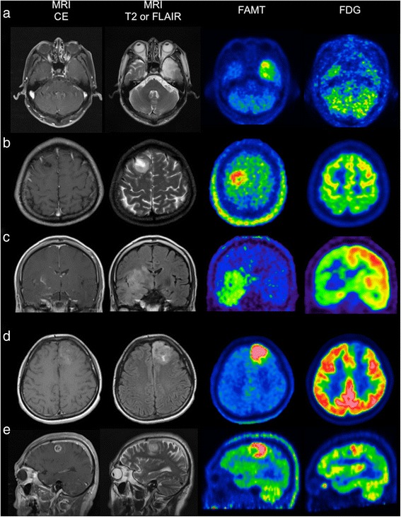
The T/N ratio of F-FAMT uptake has high positive predictive value for detection of HGGs. F-FAMT PET had higher T/N ratio, with better tumor-normal brain contrast, compared to F-FDG PET in both LGGs and HGGs. Therefore, F-FAMT is a useful radiotracer for the preoperative visualization of gliomas.
我们在38例连续的胶质瘤患者中,使用氟-18α-甲基酪氨酸(F-FAMT)正电子发射断层扫描(PET)与氟-18氟脱氧葡萄糖(F-FDG)PET相比较,研究了胶质瘤的代谢活性与组织学特征之间的关系。计算肿瘤与正常脑的比值(T/N比值),并评估T/N比值与世界卫生组织肿瘤分级或MIB-1标记指数之间的关系。使用受试者操作特征(ROC)曲线分析评估T/N比值在区分高级别胶质瘤(HGG)和低级别胶质瘤(LGG)方面的诊断价值。
F-FAMT PET的T/N比值中位数在Ⅱ级、Ⅲ级和Ⅳ级胶质瘤中分别为2.85、4.65和4.09,HGG和LGG之间存在显著差异(p = 0.006)。F-FDG PET的T/N比值(p = 0.016)和最大标准化摄取值(p = 0.033)在HGG和LGG之间也显示出显著差异。ROC分析得出F-FAMT PET的T/N比值区分HGG和LGG的最佳截断值为3.37(敏感性81%,特异性67%,准确性76%,ROC曲线下面积0.776)。阳性预测值为84%,阴性预测值为62%。在所有胶质瘤中,F-FAMT PET的T/N比值与MIB-1标记指数无相关性,而F-FDG PET的T/N比值呈正相关(r = 0.400,p = 0.013)。F-FDG和F-FAMT的T/N比值之间存在显著正相关(r = 0.454,p = 0.004),但在所有级别胶质瘤中,F-FAMT PET的T/N比值中位数显著高于F-FDG PET。
F-FAMT摄取的T/N比值对检测HGG具有较高的阳性预测价值。与F-FDG PET相比,F-FAMT PET在LGG和HGG中均具有更高的T/N比值以及更好的肿瘤与正常脑对比度。因此,F-FAMT是一种用于胶质瘤术前可视化的有用放射性示踪剂。