Murrone Domenico, Romanelli Bruno, Vella Giuseppe, Ierardi Aldo
"Di Venere" City Hospital, Unit of Neurosurgery, Bari, Italy.
"Di Venere" City Hospital, Unit of Radiology, Bari, Italy.
Int J Surg Case Rep. 2017;36:126-129. doi: 10.1016/j.ijscr.2017.05.016. Epub 2017 May 19.
Paragangliomas of filum terminale are rare benign tumors, arising from the adrenal medulla or extra-adrenal paraganglia. These lesions usually present with chronic back pain and radiculopathy and only two cases of acute neurological deficit have been reported in literature.
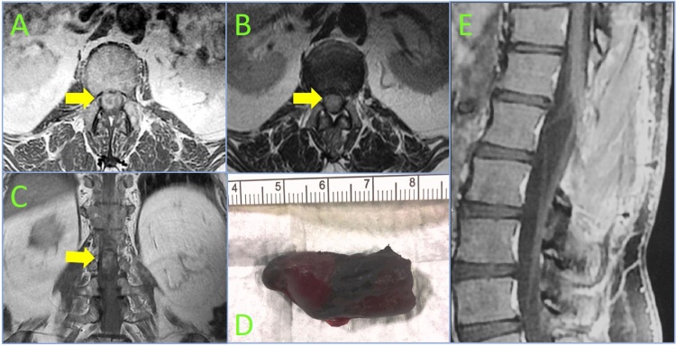
A case with an acute paraplegia and cauda equina syndrome due to an hemorrhagic paraganglioma of the filum terminale is described. Magnetic resonance imaging showed an intradural tumor extending from L1 to L2 compressing the cauda equina, with an intralesional and intradural bleed. An emergent laminectomy with total removal of the tumor was performed allowing a post-operative partial sensory recovery. Histopathological examination diagnosed paraganglioma.
Paragangliomas are solid, slow growing tumors arising from specialized neural crest cells, mostly occurring in the head and neck and rarely in cauda equina or filum terminale. MRI is gold standard radiological for diagnosis and follow-up of these lesions. They have no pathognomonic radiological and clinical features and are frequently misdiagnosed as other spinal lesions. No significant correlation was observed between the duration of symptoms and tumor dimension. Acute presentation is unusual and emergent surgical treatment is fondamental. The outcome is very good after complete excision and radiotherapical treatment is recommended after an incomplete resection.
Early radiological assessment and timely surgery are mandatory to avoid progressive neurological deficits in case of acute clinical manifestation of paraganglioma of filum terminale.
终丝副神经节瘤是一种罕见的良性肿瘤,起源于肾上腺髓质或肾上腺外副神经节。这些病变通常表现为慢性背痛和神经根病,文献中仅报道过两例急性神经功能缺损病例。
本文描述了一例因终丝出血性副神经节瘤导致急性截瘫和马尾综合征的病例。磁共振成像显示硬膜内肿瘤从L1延伸至L2,压迫马尾,肿瘤内及硬膜内有出血。急诊行椎板切除术并完整切除肿瘤,术后感觉部分恢复。组织病理学检查诊断为副神经节瘤。
副神经节瘤是起源于特殊神经嵴细胞的实性、生长缓慢的肿瘤,大多发生于头颈部,很少发生于马尾或终丝。MRI是诊断和随访这些病变的金标准影像学检查。它们没有特征性的影像学和临床特征,常被误诊为其他脊柱病变。症状持续时间与肿瘤大小之间未观察到显著相关性。急性表现不常见,急诊手术治疗至关重要。完整切除后预后非常好,不完全切除后建议行放射治疗。
对于终丝副神经节瘤急性临床表现的病例,早期影像学评估和及时手术对于避免进行性神经功能缺损是必不可少的。