Agharkar Research Institute, Developmental Biology, Pune, India.
Max Planck Institute for Heart and Lung Research, Department of Developmental Genetics, Bad Nauheim, Germany.
Sci Rep. 2017 Jun 2;7(1):2687. doi: 10.1038/s41598-017-02461-1.
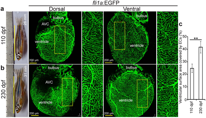
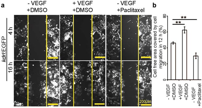
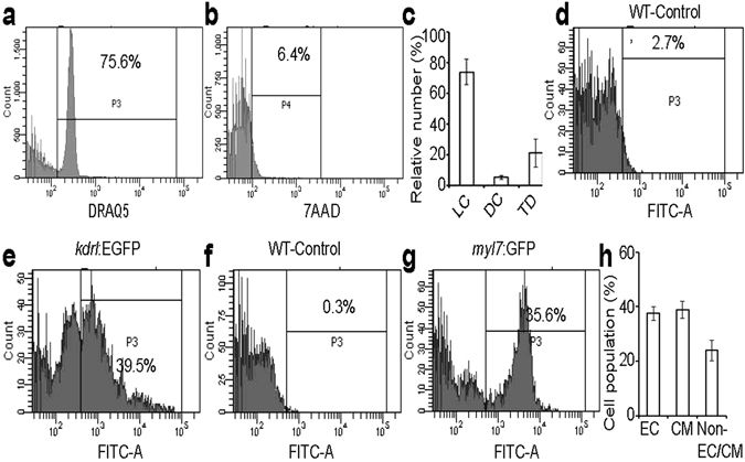
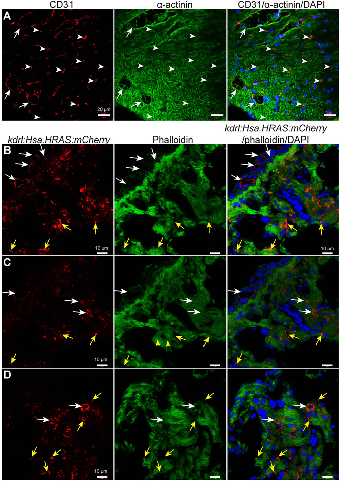
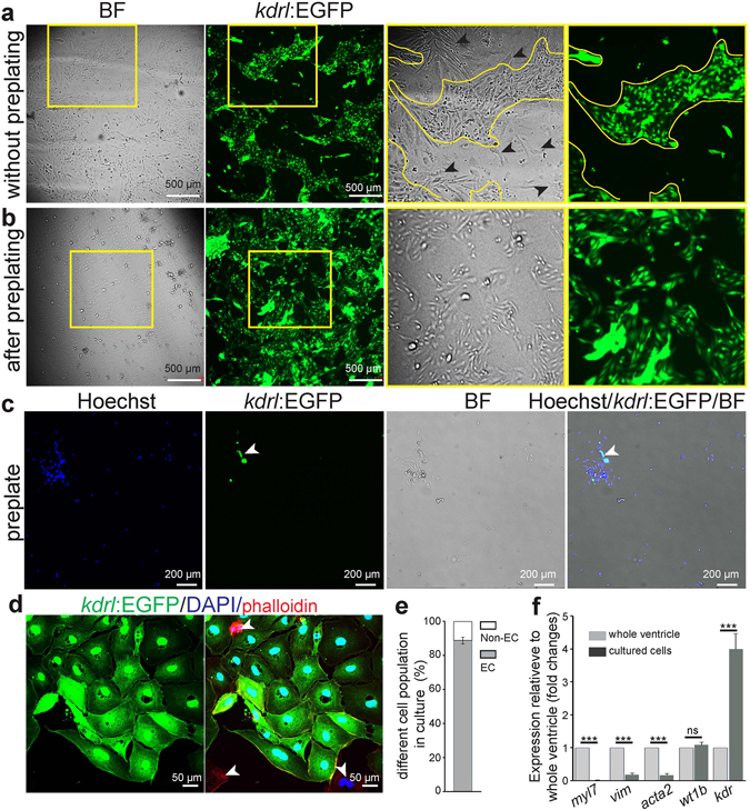
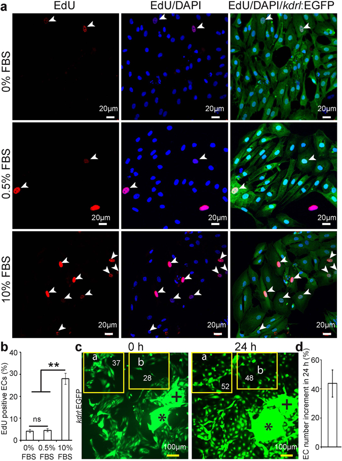
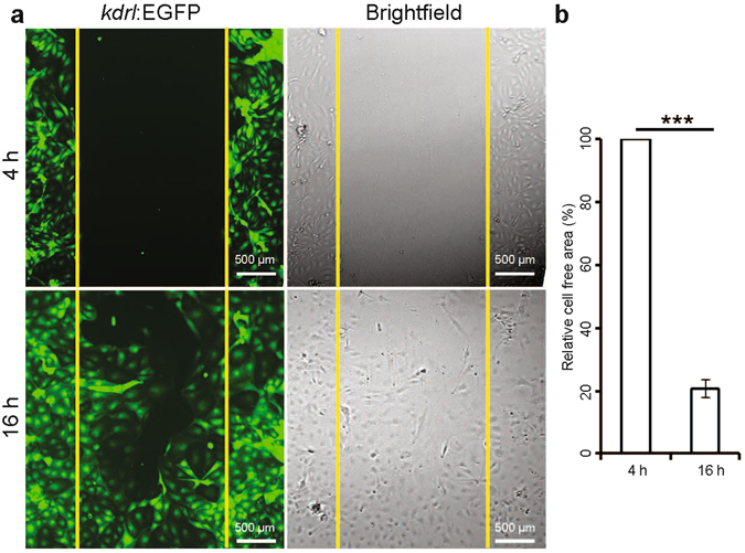
Despite our increasing understanding of zebrafish heart development and regeneration, there is limited information about the distribution of endothelial cells (ECs) in the adult zebrafish heart. Here, we investigate and compare the distribution of cardiac ECs (cECs) in adult mouse and zebrafish ventricles. Surprisingly, we find that (i) active coronary vessel growth is present in adult zebrafish, (ii) ~37 and ~39% of cells in the zebrafish heart are ECs and cardiomyocytes, respectively, a composition similar to that seen in mouse. However, we find that in zebrafish, ~36% of the ventricular tissue is covered with ECs, i.e., a substantially larger proportion than in mouse. Capitalising on the high abundance of cECs in zebrafish, we established a protocol to isolate them with high purity using fluorescent transgenic lines. Our approach eliminates side-effects due to antibody utilisation. Moreover, the isolated cECs maintained a high proliferation index even after three passages and were amenable to pharmacological treatments to study cEC migration in vitro. Such primary cultures will be a useful tool for supplementary in vitro studies on the accumulating zebrafish mutant lines as well as the screening of small molecule libraries on cardiac specific endothelial cells.
尽管我们对斑马鱼心脏发育和再生的理解不断加深,但关于成年斑马鱼心脏内皮细胞(ECs)的分布信息有限。在这里,我们研究并比较了成年小鼠和斑马鱼心室中心脏 ECs(cECs)的分布。令人惊讶的是,我们发现:(i)成年斑马鱼存在活跃的冠状血管生长;(ii)斑马鱼心脏中约有 37%和 39%的细胞分别为 ECs 和心肌细胞,这种组成与小鼠相似。然而,我们发现,在斑马鱼中,约 36%的心室组织被 ECs 覆盖,即比小鼠的比例要大得多。利用斑马鱼中大量的 cECs,我们建立了一种使用荧光转基因系以高纯度分离它们的方案。我们的方法消除了抗体使用带来的副作用。此外,分离的 cECs 在经过三次传代后仍保持较高的增殖指数,并且易于进行药理学处理以研究体外 cEC 迁移。这种原代培养将是补充在积累的斑马鱼突变系上进行体外研究以及筛选心脏特异性内皮细胞的小分子文库的有用工具。