Department of Ophthalmology, UT Southwestern Medical Center, 5323 Harry Hines Blvd., Dallas, TX 75390-9057, United States.
Department of Ophthalmology, UT Southwestern Medical Center, 5323 Harry Hines Blvd., Dallas, TX 75390-9057, United States.
Matrix Biol. 2017 Dec;64:69-80. doi: 10.1016/j.matbio.2017.06.001. Epub 2017 Jun 7.
We previously reported that fibroblasts migrating within 3-D collagen matrices move independently, whereas fibroblasts within 3-D fibrin matrices form an interconnected network. Similar networks have been identified previously during in vivo corneal wound healing. In this study, we investigate the role of fibronectin in mediating this mechanism of collective cell spreading, migration and patterning.
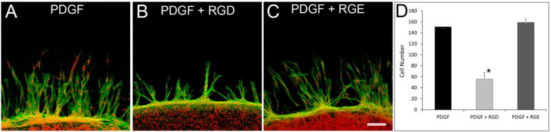
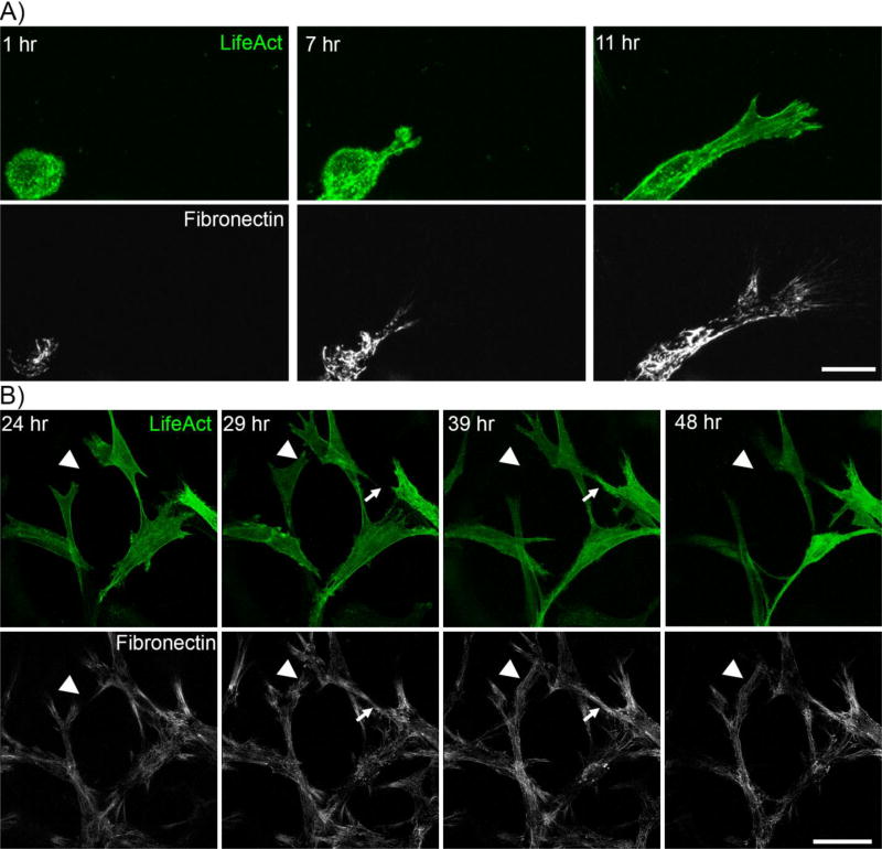
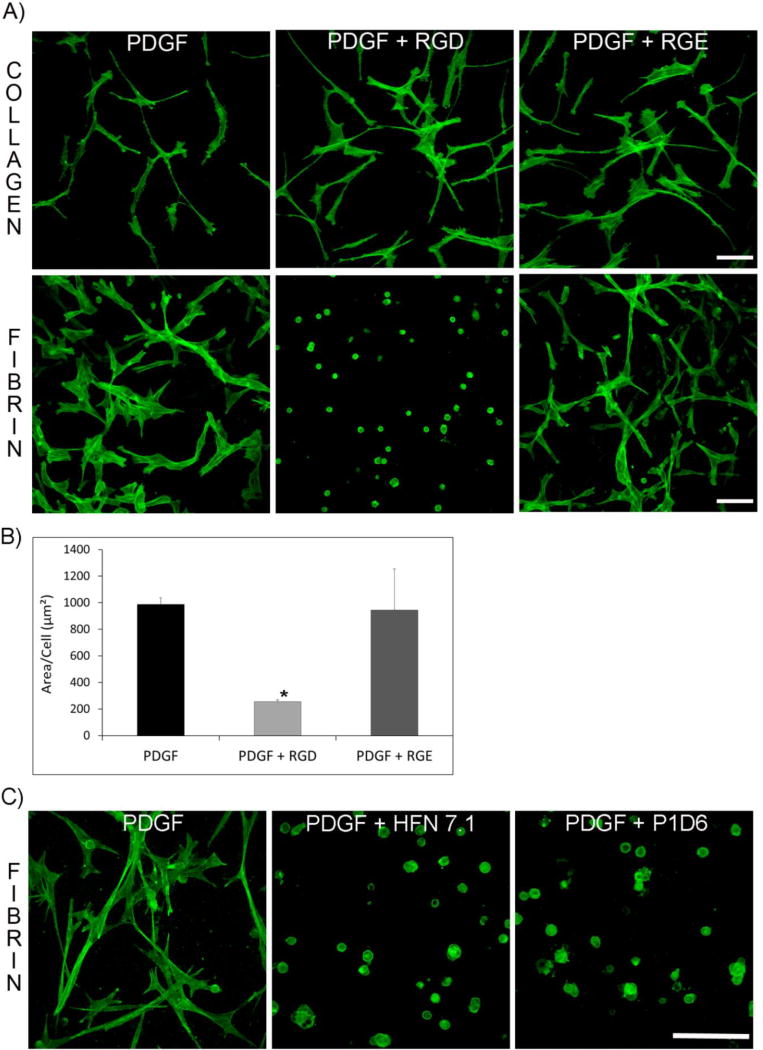
To assess cell spreading, corneal fibroblasts were plated within fibrillar collagen or fibrin matrices. To assess migration, compacted cell-populated collagen matrices were nested inside cell-free fibrin matrices. Constructs were cultured in serum-free media containing PDGF, with or without RGD peptide, anti-α5 or anti-fibronectin blocking antibodies. In some experiments, LifeAct and fluorescent fibronectin were used to allow dynamic assessment of cell-induced fibronectin reorganization. 3-D and 4-D imaging were used to assess cell mechanical behavior, connectivity, F-actin, α5 integrin and fibronectin organization.
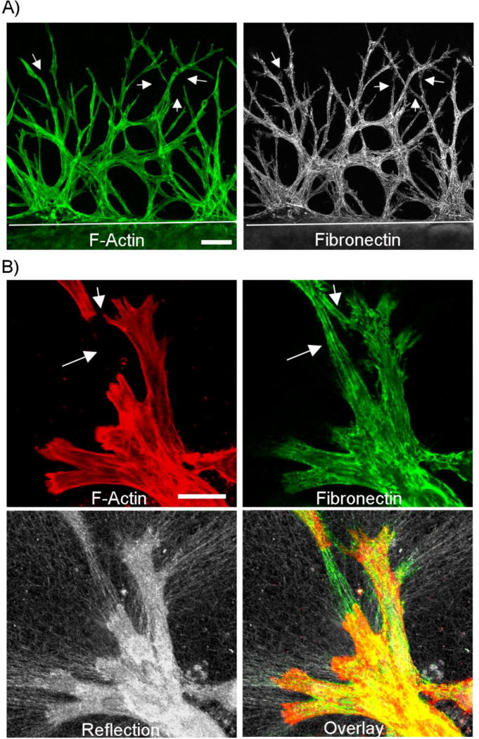
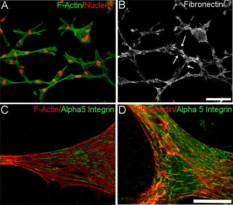
Corneal fibroblasts within 3-D fibrin matrices formed an interconnected network that was lined with cell-secreted fibronectin. Live cell imaging demonstrated that fibronectin tracks were formed at the leading edge of spreading and migrating cells. Furthermore, fibroblasts preferentially migrated through fibronectin tracks laid down by other cells. Interfering with cell-fibronectin binding with RGD, anti α5 integrin or anti fibronectin antibodies inhibited cell spreading and migration through fibrin, but did not affect cell behavior in collagen.
In this study, a novel mode of cell patterning was identified in which corneal fibroblasts secrete and attach to fibronectin via α5β1 integrin to facilitate spreading and migration within 3-D fibrin matrices, resulting in the formation of localized fibronectin tracks. Other cells use these fibronectin tracks as conduits, resulting in an interconnected cell-fibronectin network.
我们之前曾报道过,在 3D 胶原基质中迁移的成纤维细胞可以独立移动,而在 3D 纤维蛋白基质中的成纤维细胞则形成相互连接的网络。在体内角膜伤口愈合过程中也已经发现了类似的网络。在这项研究中,我们研究了纤连蛋白在介导这种细胞集体铺展、迁移和模式形成的机制中的作用。
为了评估细胞铺展,将角膜成纤维细胞种植在纤维状胶原或纤维蛋白基质中。为了评估迁移,将致密的细胞填充的胶原基质嵌套在无细胞的纤维蛋白基质内。在无血清培养基中培养构建体,其中含有 PDGF,有或没有 RGD 肽、抗α5 或抗纤连蛋白阻断抗体。在一些实验中,使用 LifeAct 和荧光纤连蛋白来允许动态评估细胞诱导的纤连蛋白重排。使用 3D 和 4D 成像来评估细胞力学行为、连通性、F-肌动蛋白、α5 整合素和纤连蛋白组织。
在 3D 纤维蛋白基质中的角膜成纤维细胞形成了一个相互连接的网络,该网络由细胞分泌的纤连蛋白排列而成。活细胞成像显示,在铺展和迁移细胞的前缘形成了纤连蛋白轨迹。此外,成纤维细胞优先通过其他细胞铺设的纤连蛋白轨迹迁移。用 RGD、抗α5 整合素或抗纤连蛋白抗体干扰细胞与纤连蛋白的结合,抑制了细胞在纤维蛋白中的铺展和迁移,但不影响细胞在胶原中的行为。
在这项研究中,确定了一种新的细胞模式形成模式,即角膜成纤维细胞通过α5β1 整合素分泌和附着纤连蛋白,以促进在 3D 纤维蛋白基质中的铺展和迁移,从而形成局部纤连蛋白轨迹。其他细胞将这些纤连蛋白轨迹用作导管,形成相互连接的细胞-纤连蛋白网络。