• 文献检索
  • 文档翻译
  • 深度研究
  • 学术资讯
  • Suppr Zotero 插件Zotero 插件
  • 邀请有礼
  • 套餐&价格
  • 历史记录
应用&插件
Suppr Zotero 插件Zotero 插件浏览器插件Mac 客户端Windows 客户端微信小程序
定价
高级版会员购买积分包购买API积分包
服务
文献检索文档翻译深度研究API 文档MCP 服务
关于我们
关于 Suppr公司介绍联系我们用户协议隐私条款
关注我们

Suppr 超能文献

核心技术专利:CN118964589B侵权必究
粤ICP备2023148730 号-1Suppr @ 2026

文献检索

告别复杂PubMed语法,用中文像聊天一样搜索,搜遍4000万医学文献。AI智能推荐,让科研检索更轻松。

立即免费搜索

文件翻译

保留排版,准确专业,支持PDF/Word/PPT等文件格式,支持 12+语言互译。

免费翻译文档

深度研究

AI帮你快速写综述,25分钟生成高质量综述,智能提取关键信息,辅助科研写作。

立即免费体验

对比增强FDG PET/CT与64层多排CT在淋巴瘤患者初始分期及治疗结束时反应评估中的多中心比较

Multicenter Comparison of Contrast-Enhanced FDG PET/CT and 64-Slice Multi-Detector-Row CT for Initial Staging and Response Evaluation at the End of Treatment in Patients With Lymphoma.

作者信息

Gómez León Nieves, Delgado-Bolton Roberto C, Del Campo Del Val Lourdes, Cabezas Beatriz, Arranz Reyes, García Marta, Cannata Jimena, González Ortega Saturnino, Pérez Sáez Mª Ángeles, López-Botet Begoña, Rodríguez-Vigil Beatriz, Mateo Marta, Colletti Patrick M, Rubello Domenico, Carreras José L

机构信息

From the *University Hospital Research Institute, Department of Radiology, University Hospital La Princesa, Madrid; †Autonomous University of Madrid, Madrid; ‡Department of Diagnostic Imaging (Radiology) and Nuclear Medicine, San Pedro Hospital and Centre for Biomedical Research of La Rioja (CIBIR), University of La Rioja, Logroño, La Rioja; Departments of §Nuclear Medicine, and ∥Radiology, University Hospital Clínico San Carlos, Madrid; ¶Department of Haematology, University Hospital la Princesa of Madrid, Madrid; Departments of **Haematology, and ††Radiology, University Hospital Fundación Jiménez Díaz, Madrid; ‡‡Department of Radiology, University Hospital Txagorritxu, Vitoria; §§Department of Haematology, University Hospital Clínico San Carlos, Madrid, Spain; ∥∥Department of Radiology, University of Southern California, Los Angeles, CA; and ¶¶Department of Nuclear Medicine, Imaging and Clinical Pathology, Santa Maria della Misericordia Hospital, Rovigo, Italy.

出版信息

Clin Nucl Med. 2017 Aug;42(8):595-602. doi: 10.1097/RLU.0000000000001718.

DOI:10.1097/RLU.0000000000001718
PMID:28604477
原文链接:https://pmc.ncbi.nlm.nih.gov/articles/PMC5502123/
Abstract

OBJECTIVES

To compare staging correctness between contrast-enhanced FDG PET/ceCT and 64-slice multi-detector-row CT (ceCT64) for initial staging and response evaluation at the end of treatment (EOT) in patients with Hodgkin lymphoma, diffuse large B cell lymphoma (DLBCL), and follicular lymphoma.

METHODS

This prospective study compared initial staging and response evaluation at EOT. One hundred eighty-one patients were randomly assigned to either ceCT64 or FDG PET/ceCT. A nuclear medicine physician and a radiologist read FDG PET/ceCT scans independently and achieved post hoc consensus, whereas another independent radiologist interpreted ceCT64 separately. The reference standard included all clinical information, all tests, and follow-up. Ethics committees of the participating centers approved the study, and all participants provided written consent.

RESULTS

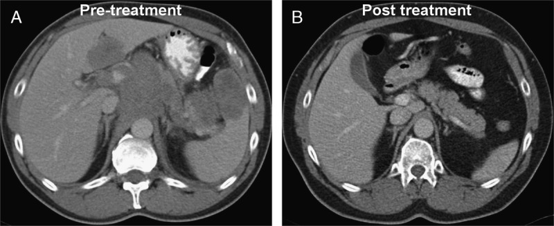
Ninety-one patients were randomized to ceCT64 and 90 to FDG PET/ceCT; 72 had Hodgkin lymphoma, 72 had DLBCL, and 37 had follicular lymphoma. There was excellent correlation between the reference standard and initial staging for both FDG PET/ceCT (κ = 0.96) and ceCT64 (κ = 0.84), although evaluation of the response at EOT was excellent only for FDG PET/ceCT (κ = 0.91).

CONCLUSIONS

Our study demonstrated satisfactory agreement between FDG PET/ceCT (κ = 0.96) and ceCT64 (κ = 0.84) in initial staging compared with the reference standard (P = 0.16). Response evaluation at EOT with FDG PET/ceCT (κ = 0.91) was superior compared with ceCT64 (κ = 0.307) (P < 0.001).

摘要

目的

比较对比增强FDG PET/ceCT与64层多排探测器CT(ceCT64)在霍奇金淋巴瘤、弥漫性大B细胞淋巴瘤(DLBCL)和滤泡性淋巴瘤患者初始分期及治疗结束时(EOT)疗效评估中的分期准确性。

方法

这项前瞻性研究比较了初始分期及EOT时的疗效评估。181例患者被随机分为ceCT64组或FDG PET/ceCT组。一名核医学医师和一名放射科医师独立解读FDG PET/ceCT扫描结果并达成事后共识,而另一名独立放射科医师单独解读ceCT64。参考标准包括所有临床信息、所有检查及随访情况。参与研究中心的伦理委员会批准了该研究,所有参与者均提供了书面同意书。

结果

91例患者被随机分配至ceCT64组,90例被分配至FDG PET/ceCT组;其中72例为霍奇金淋巴瘤,72例为DLBCL,37例为滤泡性淋巴瘤。FDG PET/ceCT(κ = 0.96)和ceCT64(κ = 0.84)的参考标准与初始分期之间均具有良好的相关性,不过仅FDG PET/ceCT对EOT时疗效的评估结果良好(κ = 0.91)。

结论

我们的研究表明,与参考标准相比,FDG PET/ceCT(κ = 0.96)和ceCT64(κ = 0.84)在初始分期方面具有令人满意的一致性(P = 0.16)。与ceCT64(κ = 0.307)相比,FDG PET/ceCT在EOT时的疗效评估更具优势(κ = 0.91)(P < 0.001)。

https://cdn.ncbi.nlm.nih.gov/pmc/blobs/7dbc/5502123/2e0ab56a363d/rlu-42-595-g006.jpg
https://cdn.ncbi.nlm.nih.gov/pmc/blobs/7dbc/5502123/ddc262442e3f/rlu-42-595-g002.jpg
https://cdn.ncbi.nlm.nih.gov/pmc/blobs/7dbc/5502123/440072092067/rlu-42-595-g004.jpg
https://cdn.ncbi.nlm.nih.gov/pmc/blobs/7dbc/5502123/e785a035e09e/rlu-42-595-g005.jpg
https://cdn.ncbi.nlm.nih.gov/pmc/blobs/7dbc/5502123/2e0ab56a363d/rlu-42-595-g006.jpg

相似文献

1
Multicenter Comparison of Contrast-Enhanced FDG PET/CT and 64-Slice Multi-Detector-Row CT for Initial Staging and Response Evaluation at the End of Treatment in Patients With Lymphoma.对比增强FDG PET/CT与64层多排CT在淋巴瘤患者初始分期及治疗结束时反应评估中的多中心比较
Clin Nucl Med. 2017 Aug;42(8):595-602. doi: 10.1097/RLU.0000000000001718.
2
Evaluation of diffuse large B-cell lymphoma patients with 64-slice multidetector computed tomography versus FDG positron emission tomography/computed tomography in initial staging and restaging after treatment.64 层多排螺旋 CT 与 FDG PET/CT 对弥漫大 B 细胞淋巴瘤初始分期和治疗后再分期的评估。
Med Clin (Barc). 2018 Oct 12;151(7):255-264. doi: 10.1016/j.medcli.2018.03.010. Epub 2018 Apr 26.
3
Diagnostic Contribution of Contrast-Enhanced CT as Compared with Unenhanced Low-Dose CT in PET/CT Staging and Treatment Response Assessment of F-FDG-Avid Lymphomas: A Prospective Study.对比增强 CT 与非增强低剂量 CT 在 PET/CT 分期和 F-FDG 摄取淋巴瘤治疗反应评估中的诊断价值:一项前瞻性研究。
J Nucl Med. 2021 Oct;62(10):1372-1379. doi: 10.2967/jnumed.120.259242. Epub 2021 Mar 12.
4
Can full-dose contrast-enhanced CT be omitted from an FDG-PET/CT staging examination in newly diagnosed FDG-avid lymphoma?在新诊断的氟代脱氧葡萄糖(FDG)摄取阳性淋巴瘤的FDG-PET/CT分期检查中,能否省略全剂量对比增强CT?
J Comput Assist Tomogr. 2014 Jul-Aug;38(4):620-5. doi: 10.1097/RCT.0000000000000067.
5
Diagnostic performance of prospective same-day 18F-FDG PET/MRI and 18F-FDG PET/CT in the staging and response assessment of lymphoma.前瞻性同日 18F-FDG PET/MRI 和 18F-FDG PET/CT 在淋巴瘤分期和疗效评估中的诊断性能。
Cancer Imaging. 2023 Jan 24;23(1):11. doi: 10.1186/s40644-023-00520-7.
6
Evaluation of diffusion-weighted MRI for pretherapeutic assessment and staging of lymphoma: results of a prospective study in 140 patients.评价弥散加权 MRI 对淋巴瘤的治疗前评估和分期的价值:140 例患者前瞻性研究结果。
Clin Cancer Res. 2014 Jun 1;20(11):2984-93. doi: 10.1158/1078-0432.CCR-13-3355. Epub 2014 Apr 2.
7
Value of FDG PET/Contrast-Enhanced CT in Initial Staging of Colorectal Cancer - Comparison with Contrast-Enhanced CT.18F-氟代脱氧葡萄糖正电子发射断层显像/增强CT在结直肠癌初始分期中的价值——与增强CT的比较
Asian Pac J Cancer Prev. 2016;17(8):4071-5.
8
Added value of pretreatment (18)F-FDG PET/CT for staging of advanced gastric cancer: Comparison with contrast-enhanced MDCT.术前(18)F-FDG PET/CT对晚期胃癌分期的附加价值:与对比增强MDCT的比较
Eur J Radiol. 2016 May;85(5):989-95. doi: 10.1016/j.ejrad.2016.03.003. Epub 2016 Mar 4.
9
Use of FDG PET/CT in identification of bone marrow involvement in diffuse large B cell lymphoma and follicular lymphoma: comparison with iliac crest bone marrow biopsy.氟代脱氧葡萄糖正电子发射断层扫描/计算机断层扫描(FDG PET/CT)在识别弥漫性大B细胞淋巴瘤和滤泡性淋巴瘤骨髓受累中的应用:与髂嵴骨髓活检的比较
Acta Radiol. 2017 Dec;58(12):1476-1484. doi: 10.1177/0284185117701305. Epub 2017 Apr 6.
10
Whole-body MRI, FDG-PET/CT, and bone marrow biopsy, for the assessment of bone marrow involvement in patients with newly diagnosed lymphoma.全身磁共振成像(MRI)、氟代脱氧葡萄糖正电子发射断层扫描/计算机断层扫描(FDG-PET/CT)以及骨髓活检,用于评估新诊断淋巴瘤患者的骨髓受累情况。
J Magn Reson Imaging. 2017 Apr;45(4):1082-1089. doi: 10.1002/jmri.25439. Epub 2016 Sep 7.

引用本文的文献

1
Whole-body MRI Versus [ 18 F]FDG PET/CT in Diagnosing and Monitoring Plasmacytomas : A Comparative Study.全身MRI与[18F]FDG PET/CT在浆细胞瘤诊断和监测中的比较研究
Clin Nucl Med. 2025 Aug 1;50(8):695-706. doi: 10.1097/RLU.0000000000005954. Epub 2025 May 16.
2
Identification of superficial invasive and indolent lymphomatous lymph nodes by multiple ultrasonographic vascular imaging.通过多种超声血管成像鉴别浅表浸润性和惰性淋巴瘤性淋巴结
Sci Rep. 2025 Mar 22;15(1):9886. doi: 10.1038/s41598-025-93545-w.
3
Predicting survival, neurotoxicity and response in B-cell lymphoma patients treated with CAR-T therapy using an imaging features-based model.

本文引用的文献

1
Clinical significance of post-treatment F-fluorodeoxyglucose uptake in cervical lymph nodes in patients with diffuse large B-cell lymphoma.弥漫性大 B 细胞淋巴瘤患者治疗后颈淋巴结 F-氟脱氧葡萄糖摄取的临床意义。
Eur Radiol. 2016 Dec;26(12):4632-4639. doi: 10.1007/s00330-016-4365-8. Epub 2016 May 18.
2
Diagnostic accuracy of FDG PET/CT for clinical evaluation at the end of treatment of HL and NHL: a comparison of the Deauville Criteria (DC) and the International Harmonization Project Criteria (IHPC).氟代脱氧葡萄糖正电子发射断层扫描/计算机断层扫描(FDG PET/CT)在霍奇金淋巴瘤(HL)和非霍奇金淋巴瘤(NHL)治疗结束时进行临床评估的诊断准确性:德奥维尔标准(DC)与国际协调项目标准(IHPC)的比较。
Eur J Nucl Med Mol Imaging. 2016 Sep;43(10):1837-48. doi: 10.1007/s00259-016-3390-9. Epub 2016 May 7.
3
使用基于成像特征的模型预测接受CAR-T疗法治疗的B细胞淋巴瘤患者的生存率、神经毒性和反应。
EJNMMI Res. 2024 Nov 20;14(1):113. doi: 10.1186/s13550-024-01172-9.
4
Prognostic value of the combination of volume, massiveness and fragmentation parameters measured on baseline FDG pet in high-burden follicular lymphoma.基线 FDG PET 上测量的体积、质量和碎裂参数的组合对高负担滤泡性淋巴瘤的预后价值。
Sci Rep. 2024 Apr 5;14(1):8033. doi: 10.1038/s41598-024-58412-0.
5
A new nomogram for assessing complete response (CR) in gastric diffuse large B-cell lymphoma (DLBCL) patients after chemotherapy.一种用于评估化疗后胃弥漫性大 B 细胞淋巴瘤 (DLBCL) 患者完全缓解 (CR) 的新诺莫图。
J Cancer Res Clin Oncol. 2023 Sep;149(12):9757-9765. doi: 10.1007/s00432-023-04862-4. Epub 2023 May 29.
6
Diffuse Large B-Cell Lymphoma Treated With R-CHOP in a Resource-Limited Setting in South Africa: A Real-World Study.南非资源有限环境下用 R-CHOP 治疗弥漫性大 B 细胞淋巴瘤的真实世界研究。
Oncologist. 2023 Sep 7;28(9):e756-e764. doi: 10.1093/oncolo/oyad069.
7
Prognostic value of genetic alterations and F-FDG PET/CT imaging features in diffuse large B cell lymphoma.弥漫性大B细胞淋巴瘤中基因改变及F-FDG PET/CT影像特征的预后价值
Am J Cancer Res. 2023 Feb 15;13(2):509-525. eCollection 2023.
8
Diagnostic performance of prospective same-day 18F-FDG PET/MRI and 18F-FDG PET/CT in the staging and response assessment of lymphoma.前瞻性同日 18F-FDG PET/MRI 和 18F-FDG PET/CT 在淋巴瘤分期和疗效评估中的诊断性能。
Cancer Imaging. 2023 Jan 24;23(1):11. doi: 10.1186/s40644-023-00520-7.
9
Limited benefit of additional contrast-enhanced CT to end-of-treatment PET/CT evaluation in patients with follicular lymphoma.在滤泡性淋巴瘤患者中,治疗结束时 PET/CT 评估中额外增强 CT 的获益有限。
Sci Rep. 2021 Sep 16;11(1):18496. doi: 10.1038/s41598-021-98081-x.
10
The characteristic computed tomography findings of pulmonary B-cell non-Hodgkin's lymphoma and their role in predicting patient survival.肺B细胞非霍奇金淋巴瘤的特征性计算机断层扫描表现及其在预测患者生存中的作用。
Quant Imaging Med Surg. 2021 Feb;11(2):772-783. doi: 10.21037/qims-20-1139.
The 2016 revision of the World Health Organization classification of lymphoid neoplasms.《世界卫生组织淋巴组织肿瘤分类(2016年修订版)》
Blood. 2016 May 19;127(20):2375-90. doi: 10.1182/blood-2016-01-643569. Epub 2016 Mar 15.
4
Detection of bone marrow involvement in newly diagnosed post-transplant lymphoproliferative disorder: (18)F-fluorodeoxyglucose positron emission tomography/computed tomography versus bone marrow biopsy.新诊断的移植后淋巴细胞增生性疾病中骨髓受累情况的检测:(18)F-氟脱氧葡萄糖正电子发射断层扫描/计算机断层扫描与骨髓活检的比较
Leuk Lymphoma. 2016 Oct;57(10):2382-8. doi: 10.3109/10428194.2016.1140162. Epub 2016 Feb 8.
5
FDG PET/CT: EANM procedure guidelines for tumour imaging: version 2.0.氟代脱氧葡萄糖正电子发射断层显像/计算机断层扫描:欧洲核医学与分子影像学会肿瘤显像程序指南:第2.0版。
Eur J Nucl Med Mol Imaging. 2015 Feb;42(2):328-54. doi: 10.1007/s00259-014-2961-x. Epub 2014 Dec 2.
6
Direct comparison of visual and quantitative bone marrow FDG-PET/CT findings with bone marrow biopsy results in diffuse large B-cell lymphoma: does bone marrow FDG-PET/CT live up to its promise?弥漫性大B细胞淋巴瘤中骨髓FDG-PET/CT视觉和定量结果与骨髓活检结果的直接比较:骨髓FDG-PET/CT是否名副其实?
Acta Radiol. 2015 Oct;56(10):1230-5. doi: 10.1177/0284185114554824. Epub 2014 Nov 11.
7
Role of imaging in the staging and response assessment of lymphoma: consensus of the International Conference on Malignant Lymphomas Imaging Working Group.影像学在淋巴瘤分期及疗效评估中的作用:恶性淋巴瘤国际会议影像工作组共识
J Clin Oncol. 2014 Sep 20;32(27):3048-58. doi: 10.1200/JCO.2013.53.5229.
8
Recommendations for initial evaluation, staging, and response assessment of Hodgkin and non-Hodgkin lymphoma: the Lugano classification.霍奇金淋巴瘤和非霍奇金淋巴瘤初始评估、分期及反应评估的建议:卢加诺分类
J Clin Oncol. 2014 Sep 20;32(27):3059-68. doi: 10.1200/JCO.2013.54.8800.
9
The predictive role of interim positron emission tomography for Hodgkin lymphoma treatment outcome is confirmed using the interpretation criteria of the Deauville five-point scale.使用多维尔五分法的解读标准证实了中期正电子发射断层扫描对霍奇金淋巴瘤治疗结果的预测作用。
Haematologica. 2014 Jun;99(6):1107-13. doi: 10.3324/haematol.2013.103218. Epub 2014 Mar 21.
10
Integrated FDG PET/CT: Utility and Applications in Clinical Oncology.一体化FDG PET/CT:在临床肿瘤学中的应用与效用
Clin Med Oncol. 2008;2:181-98. doi: 10.4137/cmo.s504. Epub 2008 Sep 19.