Hewitson Timothy D, Holt Stephen G, Tan Sven-Jean, Wigg Belinda, Samuel Chrishan S, Smith Edward R
Department of Nephrology, The Royal Melbourne Hospital, MelbourneVIC, Australia.
Department of Medicine, The Royal Melbourne Hospital, The University of Melbourne, MelbourneVIC, Australia.
Front Pharmacol. 2017 May 29;8:307. doi: 10.3389/fphar.2017.00307. eCollection 2017.
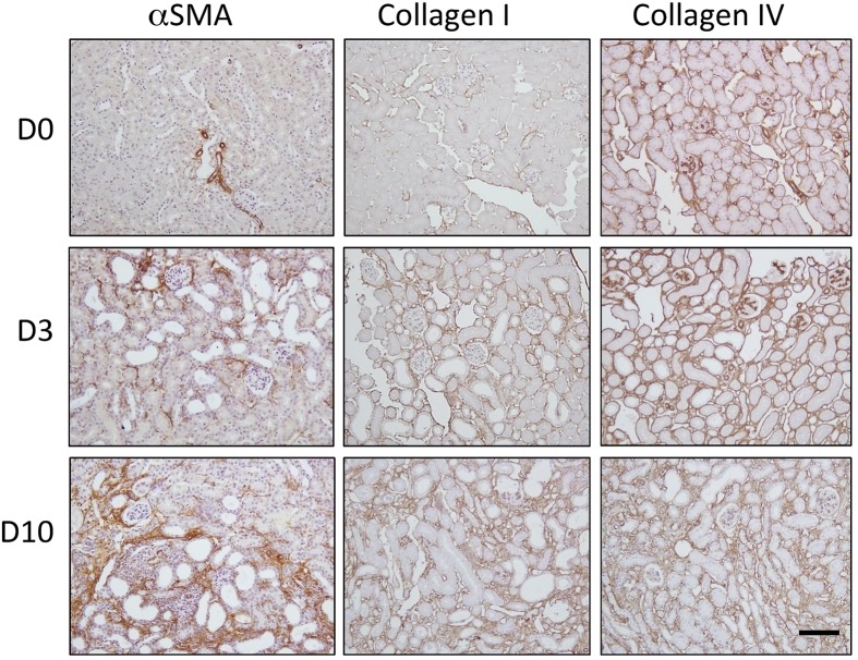
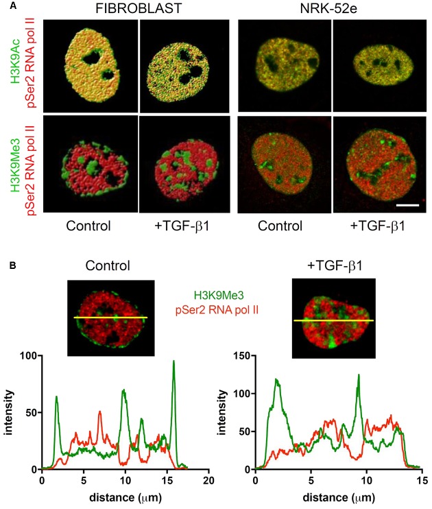
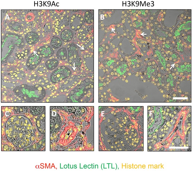
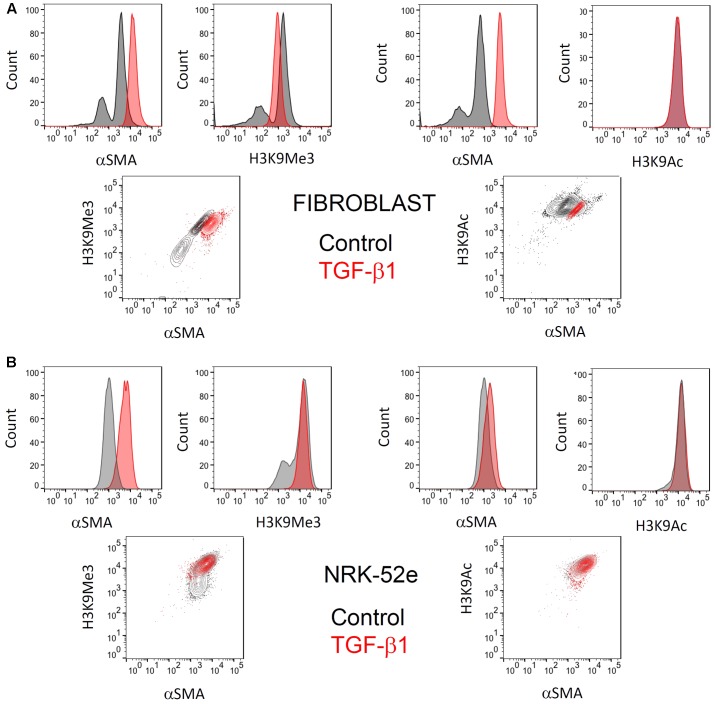
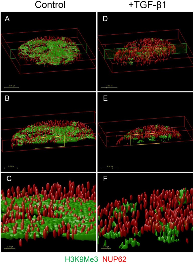
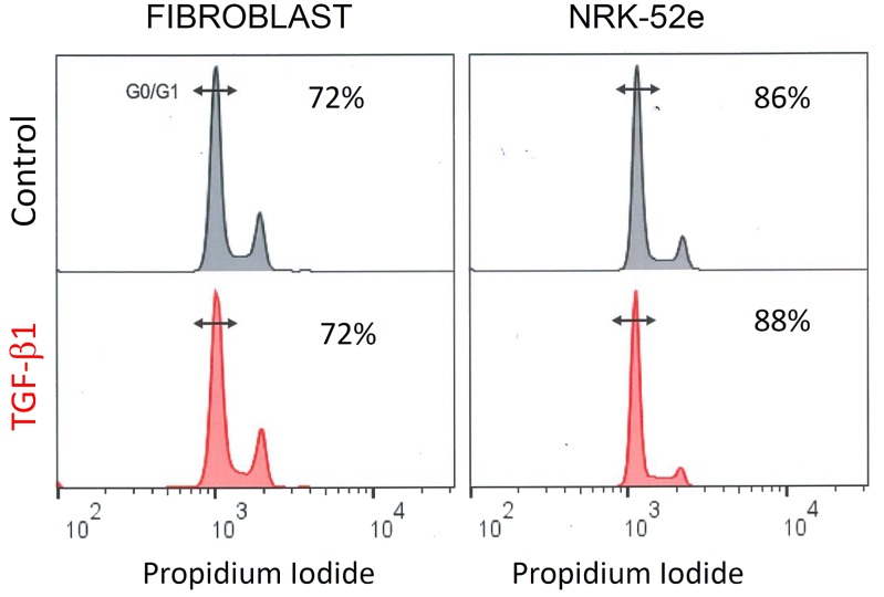
Epigenetic regulation of fibrogenesis through post-translational histone modifications (marks) may be a key determinant of progression in renal disease. In this study, we examined the distribution and acquisition of histone 3 Lysine 9 (H3K9) marks after injury and stimulation with the pro-fibrotic cytokine TGF-β1. Our focus was on their presence in activated fibroblasts (myofibroblasts) and epithelial cells (epithelial-mesenchymal transition). Immunofluorescent microscopy was used to examine global H3K9 acetylation (H3K9Ac) and tri-methylation (H3K9Me3) after unilateral ureteric obstruction (UUO) in mice. Confocal, super resolution microscopy and flow cytometry were used to determine the effect of TGF-β1 on structural arrangement of these marks, and their relationship with α-smooth muscle actin (αSMA) expression, a marker of myofibroblasts and early EMT. The number of individual histone marks was increased 10 days after UUO ( < 0.05 vs. control), with both marks clearly seen in various cell types including proximal tubules and myofibroblasts. Sub-nuclear microscopy in primary rat renal fibroblasts and a proximal tubule cell line (NRK-52e) showed that H3K9Ac was co-localized with phosphorylated-Ser2 RNA polymerase II (pRNAPol II), while H3K9Me3 was not, consistent with permissive and repressive effects on gene expression respectively. In both cell types H3K9Ac was diffusely distributed throughout the nucleus, while H3K9Me3 was found in compartments resembling the nucleolus, and in the case of the fibroblast, also juxtapositioned with the nuclear membrane. TGF-β1 had no effect on H3K9Ac marks in either cell, but resulted in a redistribution of H3K9Me3 within the fibroblast nucleus. This was unrelated to any change in mitogenesis, but was associated with increased αSMA expression. These findings highlight why it is important to consider the epigenetics of each cell individually, because whilst no overall enrichment occurred, renal myofibroblast differentiation was accompanied by distinct changes in histone mark arrangements.
通过翻译后组蛋白修饰(标记)对纤维化形成的表观遗传调控可能是肾脏疾病进展的关键决定因素。在本研究中,我们检测了在用促纤维化细胞因子转化生长因子-β1(TGF-β1)损伤和刺激后组蛋白3赖氨酸9(H3K9)标记的分布和获得情况。我们重点关注它们在活化的成纤维细胞(肌成纤维细胞)和上皮细胞(上皮-间充质转化)中的存在情况。采用免疫荧光显微镜检测小鼠单侧输尿管梗阻(UUO)后整体H3K9乙酰化(H3K9Ac)和三甲基化(H3K9Me3)情况。使用共聚焦显微镜、超分辨率显微镜和流式细胞术来确定TGF-β1对这些标记结构排列的影响,以及它们与α-平滑肌肌动蛋白(αSMA)表达的关系,αSMA是肌成纤维细胞和早期上皮-间充质转化的标志物。UUO后10天,单个组蛋白标记的数量增加(与对照组相比,P<0.05),在包括近端小管和肌成纤维细胞在内的各种细胞类型中均清晰可见这两种标记。原代大鼠肾成纤维细胞和近端小管细胞系(NRK-52e)的亚核显微镜检查显示,H3K9Ac与磷酸化的丝氨酸2 RNA聚合酶II(pRNAPol II)共定位,而H3K9Me3则不然,这分别与对基因表达的允许和抑制作用一致。在这两种细胞类型中,H3K9Ac均弥漫分布于整个细胞核,而H3K9Me3则存在于类似核仁的区域,在成纤维细胞中,还与核膜并列。TGF-β1对两种细胞中的H3K9Ac标记均无影响,但导致成纤维细胞核内H3K9Me3重新分布。这与有丝分裂发生的任何变化无关,但与αSMA表达增加有关。这些发现凸显了单独考虑每个细胞的表观遗传学为何重要,因为虽然没有发生整体富集,但肾肌成纤维细胞分化伴随着组蛋白标记排列的明显变化。