• 文献检索
  • 文档翻译
  • 深度研究
  • 学术资讯
  • Suppr Zotero 插件Zotero 插件
  • 邀请有礼
  • 套餐&价格
  • 历史记录
应用&插件
Suppr Zotero 插件Zotero 插件浏览器插件Mac 客户端Windows 客户端微信小程序
定价
高级版会员购买积分包购买API积分包
服务
文献检索文档翻译深度研究API 文档MCP 服务
关于我们
关于 Suppr公司介绍联系我们用户协议隐私条款
关注我们

Suppr 超能文献

核心技术专利:CN118964589B侵权必究
粤ICP备2023148730 号-1Suppr @ 2026

文献检索

告别复杂PubMed语法,用中文像聊天一样搜索,搜遍4000万医学文献。AI智能推荐,让科研检索更轻松。

立即免费搜索

文件翻译

保留排版,准确专业,支持PDF/Word/PPT等文件格式,支持 12+语言互译。

免费翻译文档

深度研究

AI帮你快速写综述,25分钟生成高质量综述,智能提取关键信息,辅助科研写作。

立即免费体验

基于面结构成像的二维光学显微镜数据与三维体积数据关联的新成像方法。

A novel imaging method for correlating 2D light microscopic data and 3D volume data based on block-face imaging.

机构信息

Department of Anatomy, Graduate School of Medicine, Gunma University. 39-22 Showa-machi 3-chome, Maebashi, Gunma, 371-8511, Japan.

Department of Neurosurgery, Graduate School of Medicine, Gunma University. 39-22 Showa-machi 3-chome, Maebashi, Gunma, 371-8511, Japan.

出版信息

Sci Rep. 2017 Jun 16;7(1):3645. doi: 10.1038/s41598-017-03900-9.

DOI:10.1038/s41598-017-03900-9
PMID:28623366
原文链接:https://pmc.ncbi.nlm.nih.gov/articles/PMC5473845/
Abstract

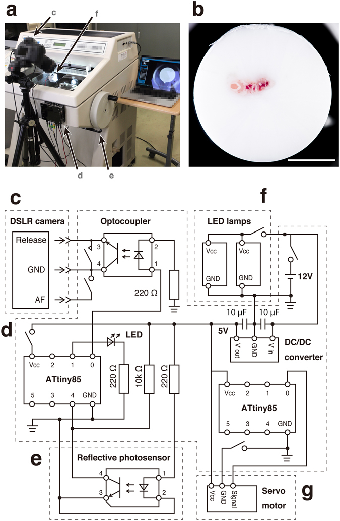
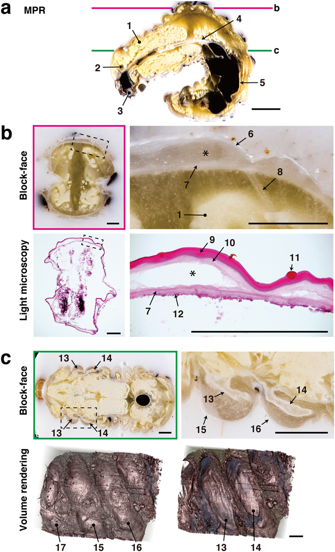
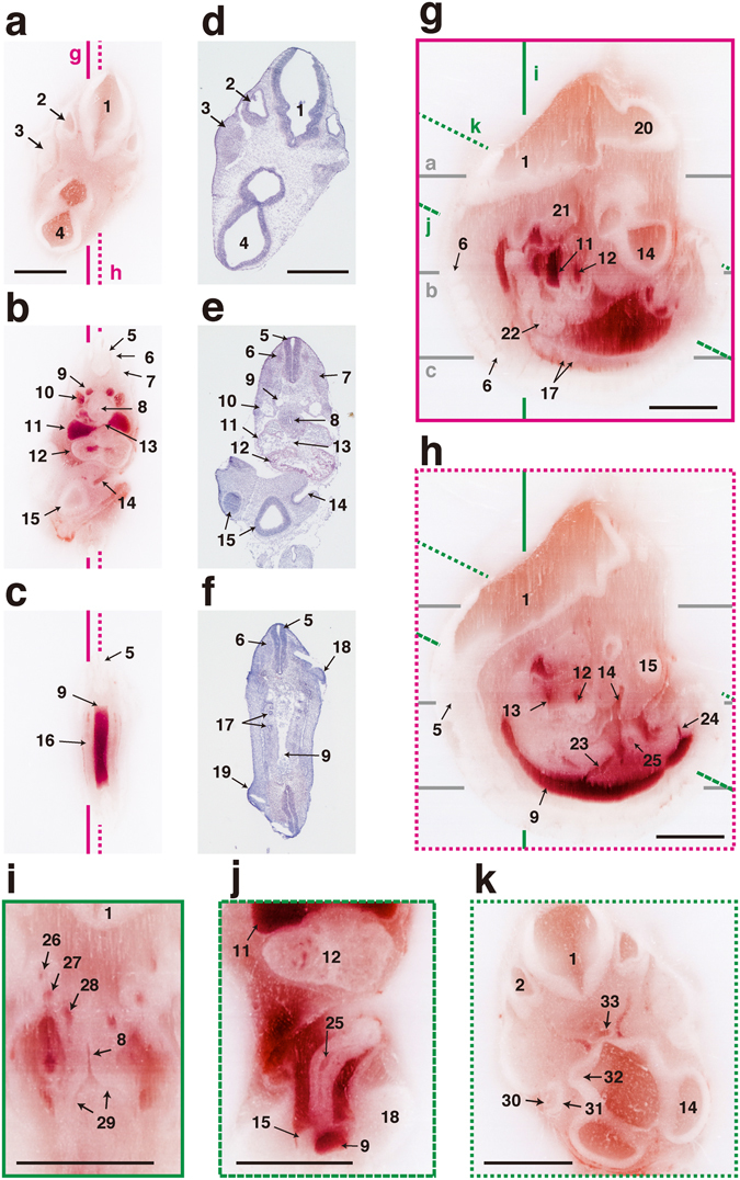
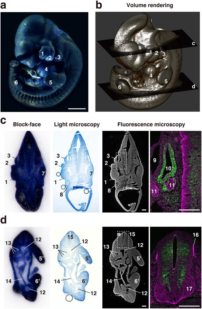
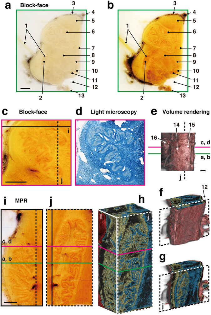
We have developed an imaging method designated as correlative light microscopy and block-face imaging (CoMBI), which contributes to improve the reliability of morphological analyses. This method can collect both the frozen sections and serial block-face images in a single specimen. The frozen section can be used for conventional light microscopic analysis to obtain 2-dimensional (2D) anatomical and molecular information, while serial block-face images can be used as 3-dimensional (3D) volume data for anatomical analysis. Thus, the sections maintain positional information in the specimen, and allows the correlation of 2D microscopic data and 3D volume data in a single specimen. The subjects can vary in size and type, and can cover most specimens encountered in biology. In addition, the required system for our method is characterized by cost-effectiveness. Here, we demonstrated the utility of CoMBI using specimens ranging in size from several millimeters to several centimeters, i.e., mouse embryos, human brainstem samples, and stag beetle larvae, and present successful correlation between the 2D light microscopic images and 3D volume data in a single specimen.

摘要

我们开发了一种成像方法,命名为相关光显微镜和块面成像(CoMBI),这有助于提高形态分析的可靠性。该方法可以在单个标本中同时收集冷冻切片和连续块面图像。冷冻切片可用于传统的光镜分析,以获得二维(2D)解剖和分子信息,而连续块面图像可用作解剖分析的三维(3D)体积数据。因此,切片在标本中保持位置信息,并允许在单个标本中对 2D 微观数据和 3D 体积数据进行关联。研究对象的大小和类型各不相同,可以涵盖生物学中遇到的大多数标本。此外,我们方法所需的系统具有成本效益。在这里,我们使用从几毫米到几厘米不等的大小的标本(即小鼠胚胎、人脑干样本和鹿角甲虫幼虫)演示了 CoMBI 的实用性,并展示了单个标本中 2D 光镜图像和 3D 体积数据之间的成功关联。

https://cdn.ncbi.nlm.nih.gov/pmc/blobs/bfac/5473845/1b8ff29796b4/41598_2017_3900_Fig5_HTML.jpg
https://cdn.ncbi.nlm.nih.gov/pmc/blobs/bfac/5473845/1e766a5a6c93/41598_2017_3900_Fig1_HTML.jpg
https://cdn.ncbi.nlm.nih.gov/pmc/blobs/bfac/5473845/07248f994970/41598_2017_3900_Fig2_HTML.jpg
https://cdn.ncbi.nlm.nih.gov/pmc/blobs/bfac/5473845/41eb267cc260/41598_2017_3900_Fig3_HTML.jpg
https://cdn.ncbi.nlm.nih.gov/pmc/blobs/bfac/5473845/313988cc4e8d/41598_2017_3900_Fig4_HTML.jpg
https://cdn.ncbi.nlm.nih.gov/pmc/blobs/bfac/5473845/1b8ff29796b4/41598_2017_3900_Fig5_HTML.jpg

相似文献

1
A novel imaging method for correlating 2D light microscopic data and 3D volume data based on block-face imaging.基于面结构成像的二维光学显微镜数据与三维体积数据关联的新成像方法。
Sci Rep. 2017 Jun 16;7(1):3645. doi: 10.1038/s41598-017-03900-9.
2
Correlative microscopy and block-face imaging (CoMBI): a 3D imaging method with wide applicability in the field of biological science.相关显微镜和块面成像(CoMBI):一种在生物科学领域具有广泛适用性的 3D 成像方法。
Anat Sci Int. 2023 Jul;98(3):353-359. doi: 10.1007/s12565-023-00705-x. Epub 2023 Feb 28.
3
Correlative microscopy and block-face imaging (CoMBI) method for both paraffin-embedded and frozen specimens.石蜡包埋和冰冻标本的相关显微镜和块面成像(CoMBI)方法。
Sci Rep. 2021 Jun 23;11(1):13108. doi: 10.1038/s41598-021-92485-5.
4
Combined method of whole mount and block-face imaging: Acquisition of 3D data of gene expression pattern from conventional in situ hybridization.整装与块面成像结合方法:从传统原位杂交获取基因表达模式的三维数据。
Dev Growth Differ. 2023 Jan;65(1):56-64. doi: 10.1111/dgd.12827. Epub 2022 Dec 13.
5
Rapid specimen preparation to improve the throughput of electron microscopic volume imaging for three-dimensional analyses of subcellular ultrastructures with serial block-face scanning electron microscopy.快速标本制备以提高用于亚细胞超微结构三维分析的电子显微镜体积成像的通量,采用连续块面扫描电子显微镜技术。
Med Mol Morphol. 2016 Sep;49(3):154-62. doi: 10.1007/s00795-016-0134-7. Epub 2016 Feb 11.
6
A rapid and efficient 2D/3D nuclear segmentation method for analysis of early mouse embryo and stem cell image data.一种快速有效的二维/三维核分割方法,用于分析早期小鼠胚胎和干细胞图像数据。
Stem Cell Reports. 2014 Mar 11;2(3):382-97. doi: 10.1016/j.stemcr.2014.01.010.
7
Exploring three-dimensional matrix-assisted laser desorption/ionization imaging mass spectrometry data: three-dimensional spatial segmentation of mouse kidney.探索三维基质辅助激光解吸/电离成像质谱数据:小鼠肾脏的三维空间分割。
Anal Chem. 2012 Jul 17;84(14):6079-87. doi: 10.1021/ac300673y. Epub 2012 Jul 5.
8
Showing their true colors: a practical approach to volume rendering from serial sections.展现真面目:一种从连续切片进行体绘制的实用方法。
BMC Dev Biol. 2010 Apr 21;10:41. doi: 10.1186/1471-213X-10-41.
9
Rapid high resolution three dimensional reconstruction of embryos with episcopic fluorescence image capture.利用落射荧光图像采集技术对胚胎进行快速高分辨率三维重建。
Birth Defects Res C Embryo Today. 2004 Sep;72(3):213-23. doi: 10.1002/bdrc.20023.
10
Three-dimensional surface texture visualization of bone tissue through epifluorescence-based serial block face imaging.通过基于落射荧光的连续块面成像实现骨组织的三维表面纹理可视化
J Microsc. 2009 Oct;236(1):52-9. doi: 10.1111/j.1365-2818.2009.03204.x.

引用本文的文献

1
The histone methyltransferase KMT2D is essential for embryo implantation via regulating precise differentiation of endometrial cells.组蛋白甲基转移酶KMT2D通过调节子宫内膜细胞的精确分化对胚胎着床至关重要。
Cell Death Discov. 2024 Aug 8;10(1):357. doi: 10.1038/s41420-024-02134-9.
2
Development of a heat-stable alkaline phosphatase reporter system for cis-regulatory analysis and its application to 3D digital imaging of Xenopus embryonic tissues.开发稳定耐热型碱性磷酸酶报告系统,用于顺式调控分析及其在爪蟾胚胎组织 3D 数字成像中的应用。
Dev Growth Differ. 2024 Apr;66(3):256-265. doi: 10.1111/dgd.12919. Epub 2024 Mar 4.
3

本文引用的文献

1
High-throughput discovery of novel developmental phenotypes.新型发育表型的高通量发现
Nature. 2016 Sep 22;537(7621):508-514. doi: 10.1038/nature19356. Epub 2016 Sep 14.
2
Light-sheet microscopy for everyone? Experience of building an OpenSPIM to study flatworm development.人人都能使用的光片显微镜?构建用于研究涡虫发育的开放式选择性平面照明显微镜的经验。
BMC Dev Biol. 2016 Jun 30;16(1):22. doi: 10.1186/s12861-016-0122-0.
3
Light-sheet microscopy imaging of a whole cleared rat brain with Thy1-GFP transgene.利用 Thy1-GFP 转基因对整个清除的大鼠脑进行光片显微镜成像。
Correlative microscopy and block-face imaging (CoMBI): a 3D imaging method with wide applicability in the field of biological science.
相关显微镜和块面成像(CoMBI):一种在生物科学领域具有广泛适用性的 3D 成像方法。
Anat Sci Int. 2023 Jul;98(3):353-359. doi: 10.1007/s12565-023-00705-x. Epub 2023 Feb 28.
4
Regenerative Polarity of the Fin Ray in Zebrafish Caudal Fin and Related Tissue Formation on the Cut Surface.斑马鱼尾鳍鳍条的再生极性及断面上相关组织的形成
J Dev Biol. 2021 Nov 19;9(4):50. doi: 10.3390/jdb9040050.
5
A Non-Invasive Approach in the Assessment of Stress Phenomena and Impairment Values in Pea Seeds Caused by Pea Weevil.一种评估豌豆象对豌豆种子造成的应激现象和损伤值的非侵入性方法。
Plants (Basel). 2021 Jul 18;10(7):1470. doi: 10.3390/plants10071470.
6
Correlative microscopy and block-face imaging (CoMBI) method for both paraffin-embedded and frozen specimens.石蜡包埋和冰冻标本的相关显微镜和块面成像(CoMBI)方法。
Sci Rep. 2021 Jun 23;11(1):13108. doi: 10.1038/s41598-021-92485-5.
7
Computational analyses decipher the primordial folding coding the 3D structure of the beetle horn.计算分析揭示了编码甲虫角 3D 结构的原始折叠密码。
Sci Rep. 2021 Jan 13;11(1):1017. doi: 10.1038/s41598-020-79757-2.
8
Neural regulation in tooth regeneration of Ambystoma mexicanum.墨西哥钝口螈牙齿再生中的神经调控。
Sci Rep. 2020 Jun 9;10(1):9323. doi: 10.1038/s41598-020-66142-2.
9
In Vivo Tracking of Tissue Engineered Constructs.组织工程构建体的体内追踪
Micromachines (Basel). 2019 Jul 16;10(7):474. doi: 10.3390/mi10070474.
10
An enhanced staining method K-B-2R staining for three-dimensional nerve reconstruction.增强型染色法 K-B-2R 用于三维神经重建。
BMC Neurosci. 2019 Jul 8;20(1):32. doi: 10.1186/s12868-019-0515-7.
Sci Rep. 2016 Jun 17;6:28209. doi: 10.1038/srep28209.
4
Three-dimensional imaging technologies: a priority for the advancement of tissue engineering and a challenge for the imaging community.三维成像技术:组织工程发展的重点及成像领域面临的挑战。
J Biophotonics. 2017 Jan;10(1):24-45. doi: 10.1002/jbio.201600049. Epub 2016 Apr 25.
5
Identification and functional analyses of sex determination genes in the sexually dimorphic stag beetle Cyclommatus metallifer.具有性别二态性的锹甲(Cyclommatus metallifer)中性别决定基因的鉴定与功能分析。
BMC Genomics. 2016 Mar 22;17:250. doi: 10.1186/s12864-016-2522-8.
6
Imaging of Neurovascular Compression Syndromes: Trigeminal Neuralgia, Hemifacial Spasm, Vestibular Paroxysmia, and Glossopharyngeal Neuralgia.神经血管压迫综合征的影像学表现:三叉神经痛、面肌痉挛、前庭阵发性眩晕和舌咽神经痛。
AJNR Am J Neuroradiol. 2016 Aug;37(8):1384-92. doi: 10.3174/ajnr.A4683. Epub 2016 Feb 18.
7
Fully automatic and robust 3D registration of serial-section microscopic images.连续切片显微镜图像的全自动且稳健的三维配准
Sci Rep. 2015 Oct 9;5:15051. doi: 10.1038/srep15051.
8
Clarifying Tissue Clearing.澄清组织透明化
Cell. 2015 Jul 16;162(2):246-257. doi: 10.1016/j.cell.2015.06.067.
9
Correlative microscopy.相关显微镜检查
Arch Biochem Biophys. 2015 Sep 1;581:98-110. doi: 10.1016/j.abb.2015.05.017. Epub 2015 Jun 10.
10
From Animaculum to single molecules: 300 years of the light microscope.从微生物到单分子:光学显微镜的三百年
Open Biol. 2015 Apr;5(4):150019. doi: 10.1098/rsob.150019.