Pellegrini Giovanni, Siwowska Klaudia, Haller Stephanie, Antoine Daniel J, Schibli Roger, Kipar Anja, Müller Cristina
Laboratory for Animal Model Pathology (LAMP), Institute of Veterinary Pathology, Vetsuisse Faculty, University of Zurich, 8057 Zurich, Switzerland.
Center for Radiopharmaceutical Sciences ETH-PSI-USZ, Paul Scherrer Institut, 5232 Villigen-PSI, Switzerland.
Pharmaceuticals (Basel). 2017 Jun 21;10(2):57. doi: 10.3390/ph10020057.
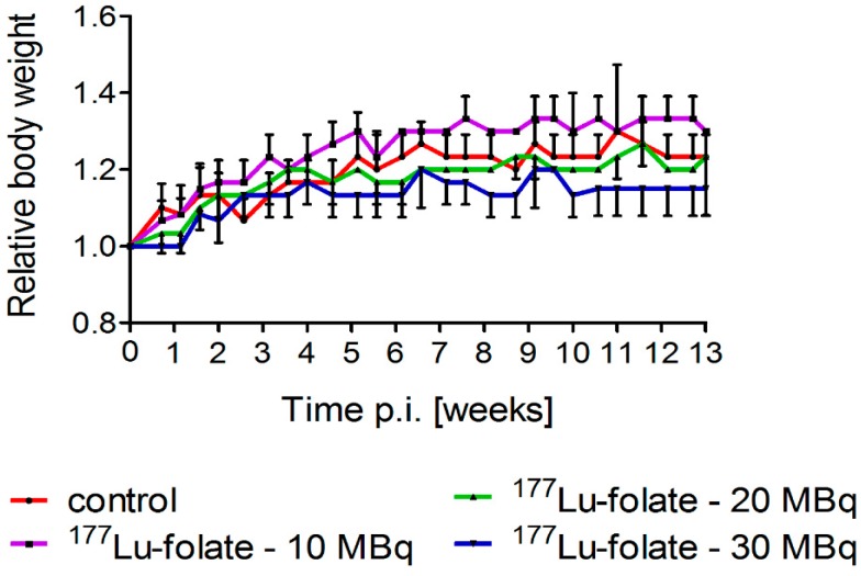
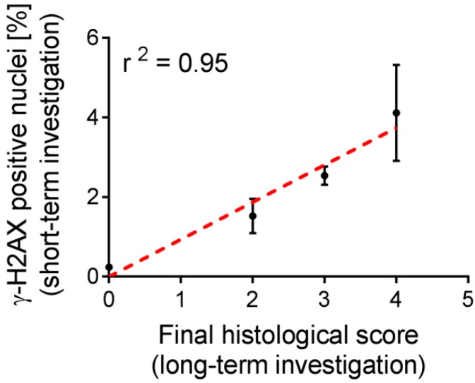
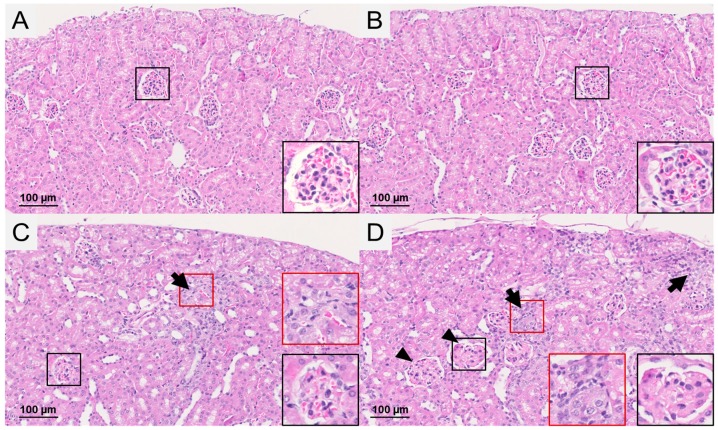
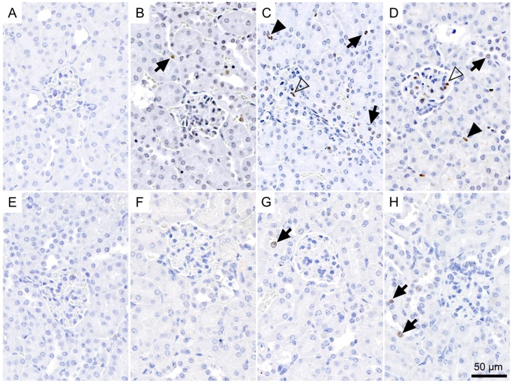
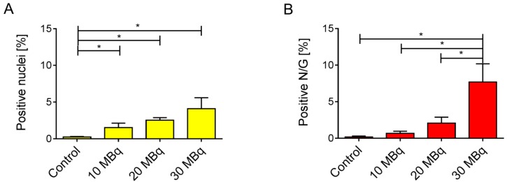
Folate receptor (FR)-targeted radionuclide therapy using folate radioconjugates is of interest due to the expression of the FR in a variety of tumor types. The high renal accumulation of radiofolates presents, however, a risk of radionephropathy. A potential option to address this challenge would be to use radioprotectants, such as amifostine. Methods for early detection of kidney damage that-in this case-cannot be predicted based on dose estimations, would facilitate the development of novel therapies. The aim of this study was, therefore, to assess potentially changing levels of plasma and urine biomarkers and to determine DNA damage at an early stage after radiofolate application. The identification of an early indicator for renal damage in mice would be useful since histological changes become apparent only several months after treatment. Mice were injected with different quantities of Lu-folate (10 MBq, 20 MBq and 30 MBq), resulting in mean absorbed kidney doses of ~23 Gy, ~46 Gy and ~69 Gy, respectively, followed by euthanasia two weeks (>85% of the mean renal radiation dose absorbed) or three months later. Whereas all investigated biomarkers remained unchanged, the number of γ-H2AX-positive nuclei in the renal cortex showed an evident dose-dependent increase as compared to control values two weeks after treatment. Comparison with the extent of kidney injury determined by histological changes five to eight months after administration of the same Lu-folate activities suggested that the quantitative assessment of double-strand breaks can be used as a biological indicator for long-term radiation effects in the kidneys. This method may, thus, enable faster assessment of radiopharmaceuticals and protective measures by preventing logistically challenging long-term investigations to detect kidney damage.
由于叶酸受体(FR)在多种肿瘤类型中表达,使用叶酸放射性缀合物进行叶酸受体靶向放射性核素治疗备受关注。然而,放射性叶酸在肾脏中的高蓄积存在放射性肾病的风险。应对这一挑战的一个潜在选择是使用辐射防护剂,如氨磷汀。对于这种无法基于剂量估计进行预测的肾脏损伤,早期检测方法将有助于新型疗法的开发。因此,本研究的目的是评估放射性叶酸应用后早期血浆和尿液生物标志物水平的潜在变化,并确定DNA损伤情况。确定小鼠肾脏损伤的早期指标将很有用,因为组织学变化在治疗后几个月才会明显显现。给小鼠注射不同剂量的镥 - 叶酸(10 MBq、20 MBq和30 MBq),分别导致平均肾脏吸收剂量约为23 Gy、约46 Gy和约69 Gy,然后在两周(>平均肾脏吸收辐射剂量的85%)或三个月后实施安乐死。尽管所有研究的生物标志物均未发生变化,但与对照组相比,治疗两周后肾皮质中γ - H2AX阳性细胞核的数量呈现出明显的剂量依赖性增加。将其与在给予相同镥 - 叶酸活性五至八个月后通过组织学变化确定的肾脏损伤程度进行比较表明,双链断裂的定量评估可作为肾脏长期辐射效应的生物学指标。因此,这种方法可以通过避免为检测肾脏损伤而进行的后勤保障要求高的长期研究,实现对放射性药物和防护措施的更快评估。