Almeida Vanessa Camillo, Pinheiro Lucas Rodrigues, Salineiro Fernanda Cristina Sales, Mendes Fausto Medeiros, Neto João Batista César, Cavalcanti Marcelo Gusmão Paraíso, Pannuti Cláudio Mendes
Department of Periodontology, School of Dentistry, University of São Paulo, Av. Prof. Lineu Prestes, 2227, São Paulo, SP, 05508-000, Brazil.
Department of Radiology, School of Dentistry, University of São Paulo, Av. Prof. Lineu Prestes, 2227, São Paulo, SP, 05508-000, Brazil.
BMC Oral Health. 2017 Jun 21;17(1):100. doi: 10.1186/s12903-017-0390-5.
Cone beam computed tomography (CBCT) has been largely used in dentistry. Nevertheless, there is lack of evidence regarding CBCT accuracy in the diagnosis of early periodontal lesions as well as the correlation between accuracy and lesion size. The aim of this study was to evaluate accuracy of CBCT and conventional intraoral radiographs in detecting different-sized interproximal bone lesions created in pig mandibles. The hypothesis was that CBCT accuracy would be superior to radiographs in detecting incipient bone lesions.
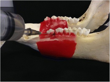
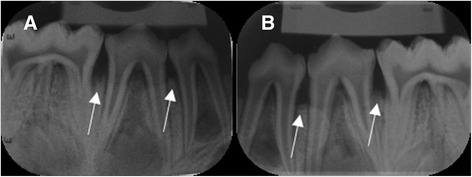
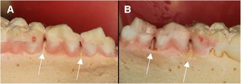
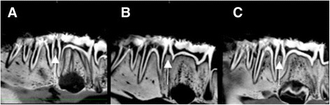
Twenty swine dry mandibles were used, totalizing 80 experimental sites. Four groups were created according to exposure time to perchloric acid 70-72%: controls (no exposure), 2-hour exposure, 4-hour exposure, and 6-hour exposure. Standardized CBCT and conventional intraoral radiographs were taken and analyzed by two trained radiologists. The presence of lesions in the dry mandible was considered the gold standard. Sensitivity, specificity, and accuracy in detecting different-sized bone lesions were calculated for CBCT and intraoral radiographs.
Accuracy of CBCT ranged from 0.762 to 0.825 and accuracy of periapical radiography ranged from 0.700 to 0.813, according to examiner and time of acid exposure. Inter-examiner agreement varied from slight to fair, whereas intra-examiner agreement varied from moderate to substantial.
CBCT performance was not superior to that provided by conventional intraoral radiographs in the detection of interproximal bone loss.
锥形束计算机断层扫描(CBCT)已在牙科领域广泛应用。然而,关于CBCT在早期牙周病变诊断中的准确性以及准确性与病变大小之间的相关性,目前仍缺乏证据。本研究的目的是评估CBCT和传统口腔内X光片在检测猪下颌骨中不同大小的邻间骨病变方面的准确性。假设是在检测初期骨病变时,CBCT的准确性将优于X光片。
使用20个猪的干燥下颌骨,共有80个实验部位。根据暴露于70 - 72%高氯酸的时间创建四组:对照组(未暴露)、2小时暴露组、4小时暴露组和6小时暴露组。由两名经过培训的放射科医生拍摄并分析标准化的CBCT和传统口腔内X光片。将干燥下颌骨中病变的存在视为金标准。计算CBCT和口腔内X光片在检测不同大小骨病变时的敏感性、特异性和准确性。
根据检查者和酸暴露时间,CBCT的准确性范围为0.762至0.825,根尖片的准确性范围为0.700至0.813。检查者间的一致性从轻微到一般,而检查者内的一致性从中度到高度。
在检测邻间骨丢失方面,CBCT的表现并不优于传统口腔内X光片。