Division of Nephrology, Department of Medicine, University of Alabama at Birmingham, Birmingham, AL, USA.
Department of Biostatistics, Ryals School of Public Health, University of Alabama at Birmingham, Birmingham, AL, USA.
Acta Physiol (Oxf). 2018 Feb;222(2). doi: 10.1111/apha.12913. Epub 2017 Jul 13.
Sphingosine-1-phosphate (S1P) influences resistance vessel function and is implicated in renal pathological processes. Previous studies revealed that S1P evoked potent vasoconstriction of the pre-glomerular microvasculature, but the underlying mechanisms remain incompletely defined. We postulated that S1P-mediated pre-glomerular microvascular vasoconstriction involves activation of voltage-dependent L-type calcium channels (L-VDCC) and the rho/rho kinase pathway.
Afferent arteriolar reactivity was assessed in vitro using the blood-perfused rat juxtamedullary nephron preparation, and diameter was measured during exposure to physiological and pharmacological agents.
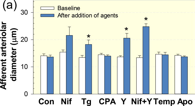
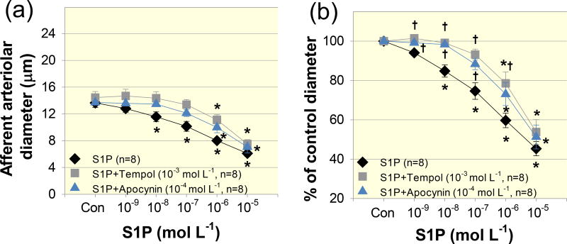
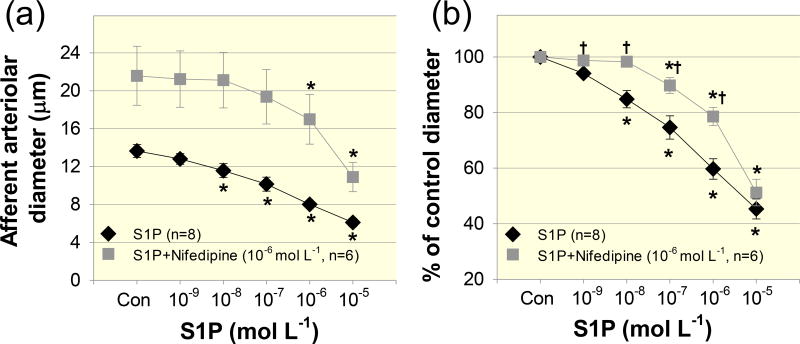
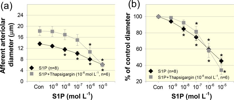
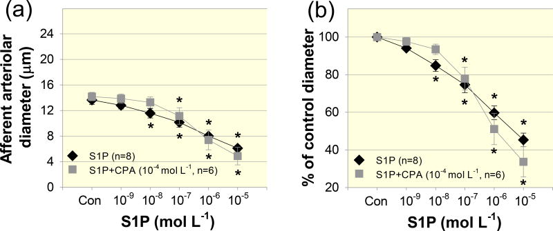
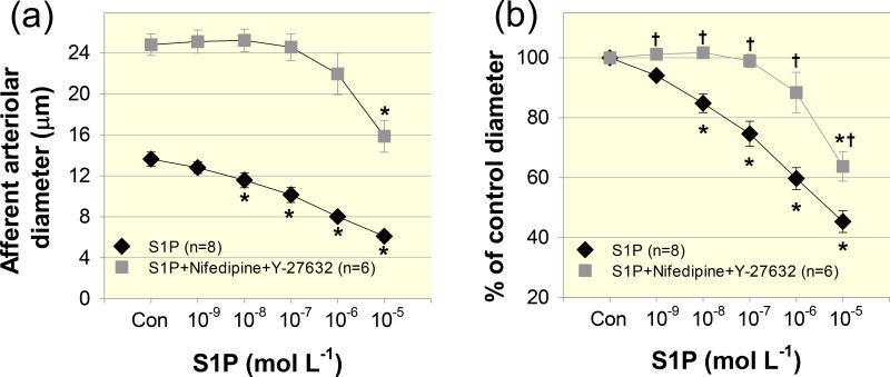
Exogenous S1P (10 -10 mol L ) evoked concentration-dependent vasoconstriction of afferent arterioles. Superfusion with nifedipine, a L-VDCC blocker, increased arteriolar diameter by 39 ± 18% of baseline and significantly attenuated the S1P-induced vasoconstriction. Superfusion with the rho kinase inhibitor, Y-27632, increased diameter by 60 ± 12% of baseline and also significantly blunted vasoconstriction by S1P. Combined nifedipine and Y-27632 treatment significantly inhibited S1P-induced vasoconstriction over the entire concentration range tested. In contrast, depletion of intracellular Ca stores with the Ca -ATPase inhibitors, thapsigargin or cyclopiazonic acid, did not alter the S1P-mediated vasoconstrictor profile. Scavenging reactive oxygen species (ROS) or inhibition of nicotinamide adenine dinucleotide phosphate oxidase activity significantly attenuated S1P-mediated vasoconstriction.
Exogenous S1P elicits potent vasoconstriction of rat afferent arterioles. These data also demonstrate that S1P-mediated pre-glomerular vasoconstriction involves activation of L-VDCC, the rho/rho kinase pathway and ROS. Mobilization of Ca from intracellular stores is not required for S1P-mediated vasoconstriction. These studies reveal a potential role for S1P in the modulation of renal microvascular tone.
鞘氨醇-1-磷酸(S1P)影响阻力血管的功能,并与肾脏病理过程有关。先前的研究表明,S1P 可引起肾小球前微血管强烈收缩,但潜在机制尚不完全明确。我们推测 S1P 介导的肾小球前微血管收缩涉及电压依赖性 L 型钙通道(L-VDCC)和 rho/ rho 激酶通路的激活。
使用血液灌注的大鼠近髓肾单位制备物在体外评估入球小动脉的反应性,并在暴露于生理和药理学试剂时测量直径。
外源性 S1P(10-10 mol L)可引起入球小动脉浓度依赖性收缩。用硝苯地平(L-VDCC 阻断剂)灌流可使小动脉直径增加 39±18%基础值,并显著减弱 S1P 引起的收缩。用 rho 激酶抑制剂 Y-27632 灌流可使直径增加 60±12%基础值,并显著减弱 S1P 引起的收缩。硝苯地平与 Y-27632 联合处理可显著抑制整个测试浓度范围内 S1P 引起的收缩。相比之下,用钙 -ATP 酶抑制剂 thapsigargin 或环匹阿尼酸耗尽细胞内钙库不会改变 S1P 介导的血管收缩特征。清除活性氧(ROS)或抑制烟酰胺腺嘌呤二核苷酸磷酸氧化酶活性可显著减弱 S1P 介导的血管收缩。
外源性 S1P 引起大鼠入球小动脉强烈收缩。这些数据还表明,S1P 介导的肾小球前血管收缩涉及 L-VDCC、rho/ rho 激酶通路和 ROS 的激活。细胞内钙库中钙的动员不是 S1P 介导的血管收缩所必需的。这些研究揭示了 S1P 在调节肾脏微血管张力中的潜在作用。