Crawford Melissa, Leclerc Valerie, Dagnino Lina
Dept. of Physiology and Pharmacology, Children's Health Research Institute and Lawson Health Research Institute, The University of Western Ontario, London, Ontario N6A 5C1, Canada.
Dept. of Physiology and Pharmacology, Children's Health Research Institute and Lawson Health Research Institute, The University of Western Ontario, London, Ontario N6A 5C1, Canada
Biol Open. 2017 Aug 15;6(8):1219-1228. doi: 10.1242/bio.025833.
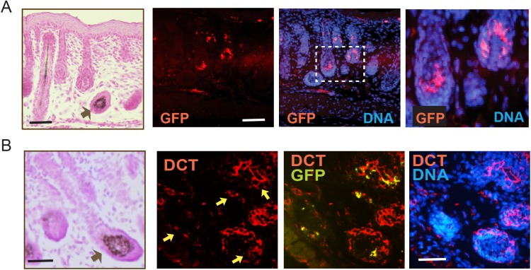
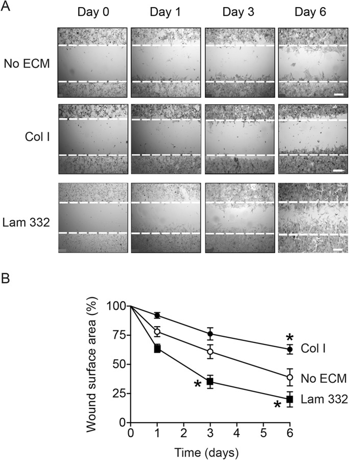
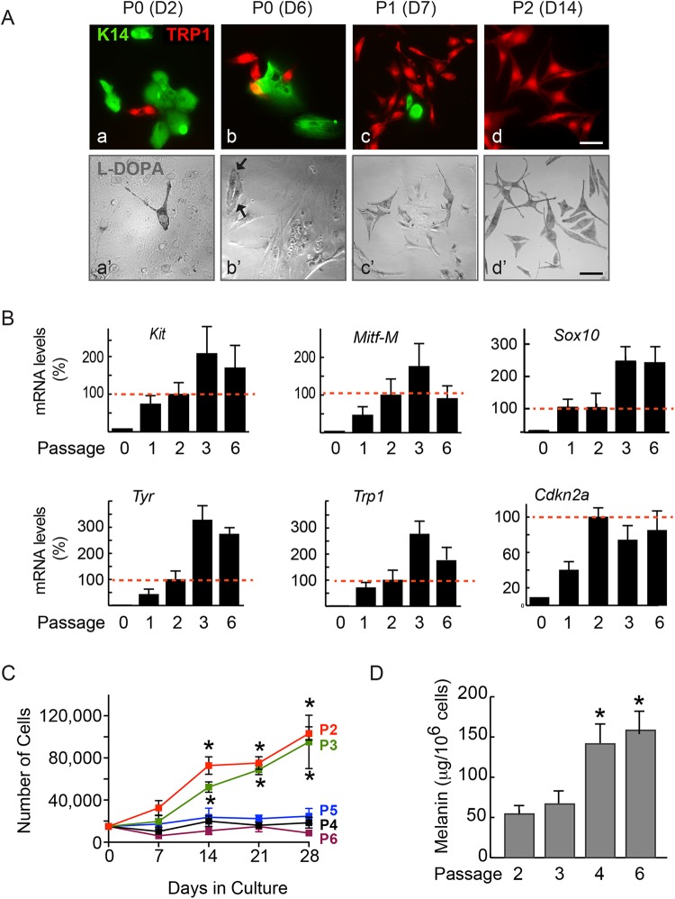
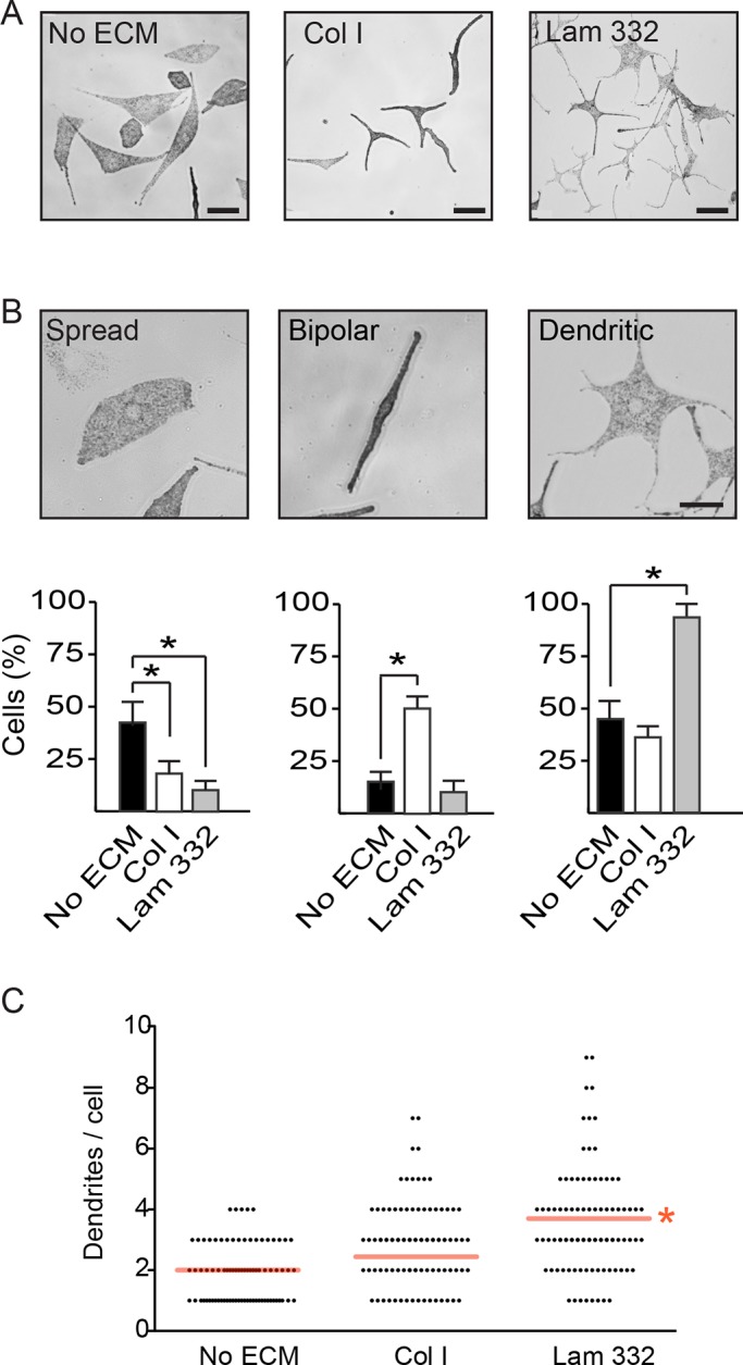
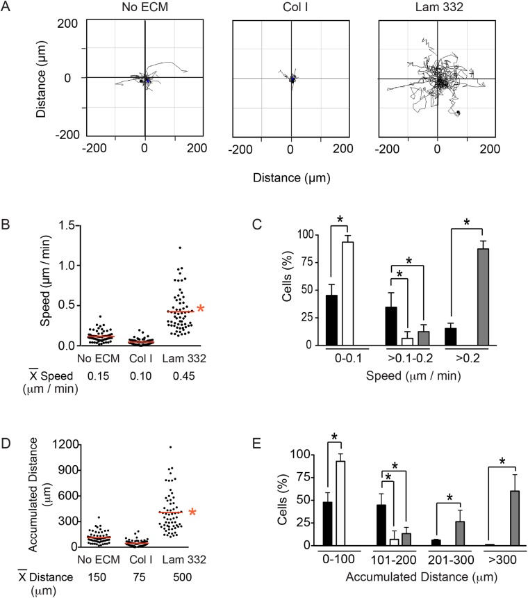
Alterations in melanocytic lineage cells give rise to a plethora of distinct human diseases, including neurocristopathies, cutaneous pigmentation disorders, loss of vision and hearing, and melanoma. Understanding the ontogeny and biology of melanocytic cells, as well as how they interact with their surrounding environment, are key steps in the development of therapies for diseases that involve this cell lineage. Efforts to culture and characterize primary melanocytes from normal or genetically engineered mouse models have at times yielded contrasting observations. This is due, in part, to differences in the conditions used to isolate, purify and culture these cells in individual studies. By breeding ROSA and mice, we generated animals in which melanocytic lineage cells are identified through expression of green fluorescent protein. We also used defined conditions to systematically investigate the proliferation and migration responses of primary melanocytes on various extracellular matrix (ECM) substrates. Under our culture conditions, mouse melanocytes exhibit doubling times in the range of 10 days, and retain exponential proliferative capacity for 50-60 days. In culture, these melanocytes showed distinct responses to different ECM substrates. Specifically, laminin-332 promoted cell spreading, formation of dendrites, random motility and directional migration. In contrast, low or intermediate concentrations of collagen I promoted adhesion and acquisition of a bipolar morphology, and interfered with melanocyte forward movements. Our systematic evaluation of primary melanocyte responses emphasizes the importance of clearly defining culture conditions for these cells. This, in turn, is essential for the interpretation of melanocyte responses to extracellular cues and to understand the molecular basis of disorders involving the melanocytic cell lineage.
黑素细胞系细胞的改变会引发大量不同的人类疾病,包括神经嵴病、皮肤色素沉着障碍、视力和听力丧失以及黑色素瘤。了解黑素细胞的个体发生和生物学特性,以及它们如何与周围环境相互作用,是开发涉及该细胞系疾病治疗方法的关键步骤。从正常或基因工程小鼠模型中培养和鉴定原代黑素细胞的努力有时会得出相互矛盾的观察结果。这部分是由于在个别研究中用于分离、纯化和培养这些细胞的条件存在差异。通过培育ROSA和 小鼠,我们培育出了通过绿色荧光蛋白表达来鉴定黑素细胞系细胞的动物。我们还使用特定条件系统地研究了原代黑素细胞在各种细胞外基质(ECM)底物上的增殖和迁移反应。在我们的培养条件下,小鼠黑素细胞的倍增时间在10天左右,并在50 - 60天内保持指数增殖能力。在培养中,这些黑素细胞对不同的ECM底物表现出不同的反应。具体而言,层粘连蛋白-332促进细胞铺展、树突形成、随机运动和定向迁移。相比之下,低浓度或中等浓度的I型胶原促进细胞黏附并获得双极形态,并干扰黑素细胞向前运动。我们对原代黑素细胞反应的系统评估强调了明确界定这些细胞培养条件的重要性。反过来,这对于解释黑素细胞对细胞外信号的反应以及理解涉及黑素细胞系的疾病的分子基础至关重要。