• 文献检索
  • 文档翻译
  • 深度研究
  • 学术资讯
  • Suppr Zotero 插件Zotero 插件
  • 邀请有礼
  • 套餐&价格
  • 历史记录
应用&插件
Suppr Zotero 插件Zotero 插件浏览器插件Mac 客户端Windows 客户端微信小程序
定价
高级版会员购买积分包购买API积分包
服务
文献检索文档翻译深度研究API 文档MCP 服务
关于我们
关于 Suppr公司介绍联系我们用户协议隐私条款
关注我们

Suppr 超能文献

核心技术专利:CN118964589B侵权必究
粤ICP备2023148730 号-1Suppr @ 2026

文献检索

告别复杂PubMed语法,用中文像聊天一样搜索,搜遍4000万医学文献。AI智能推荐,让科研检索更轻松。

立即免费搜索

文件翻译

保留排版,准确专业,支持PDF/Word/PPT等文件格式,支持 12+语言互译。

免费翻译文档

深度研究

AI帮你快速写综述,25分钟生成高质量综述,智能提取关键信息,辅助科研写作。

立即免费体验

结缔组织病相关间质性肺疾病中的放射学胸膜实质纤维弹性组织增生样病变

Radiologic pleuroparenchymal fibroelastosis-like lesion in connective tissue disease-related interstitial lung disease.

作者信息

Enomoto Yasunori, Nakamura Yutaro, Colby Thomas V, Johkoh Takeshi, Sumikawa Hiromitsu, Nishimoto Koji, Yoshimura Katsuhiro, Matsushima Sayomi, Oyama Yoshiyuki, Hozumi Hironao, Kono Masato, Fujisawa Tomoyuki, Enomoto Noriyuki, Inui Naoki, Iwashita Toshihide, Suda Takafumi

机构信息

Second Division, Department of Internal Medicine, Hamamatsu University School of Medicine, Shizuoka, Japan.

Department of Regenerative and Infectious Pathology, Hamamatsu University School of Medicine, Shizuoka, Japan.

出版信息

PLoS One. 2017 Jun 30;12(6):e0180283. doi: 10.1371/journal.pone.0180283. eCollection 2017.

DOI:10.1371/journal.pone.0180283
PMID:28666014
原文链接:https://pmc.ncbi.nlm.nih.gov/articles/PMC5493376/
Abstract

BACKGROUND

Radiologic pleuroparenchymal fibroelastosis (PPFE)-like lesion including pulmonary apical cap can be occasionally observed in clinical settings. However, the significance of radiologic PPFE-like lesion is unclear in connective tissue disease (CTD)-related interstitial lung disease (ILD).

MATERIALS AND METHODS

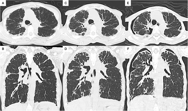
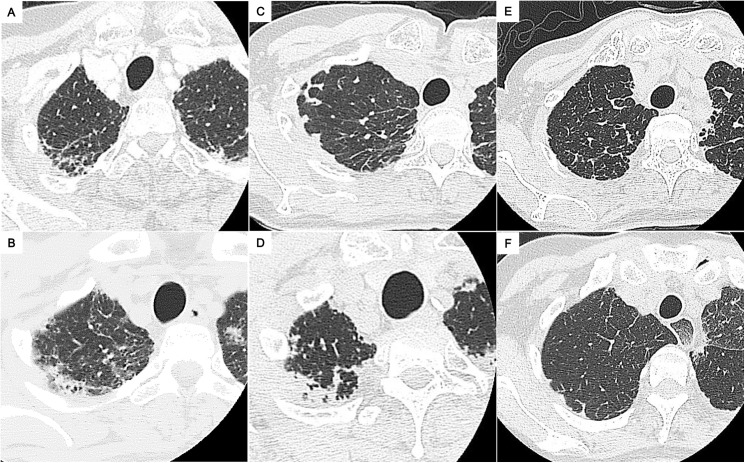
A total of 113 patients with CTD-related ILD were enrolled and assessed for radiologic PPFE-like lesion, which was defined as bilateral, upper lobe, and subpleural dense consolidations with or without pleural thickening on chest high-resolution computed tomography. The clinical, radiologic, and pathologic characteristics were evaluated.

RESULTS

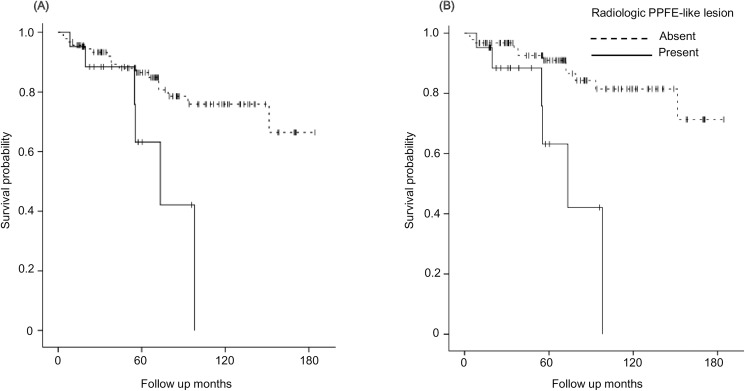
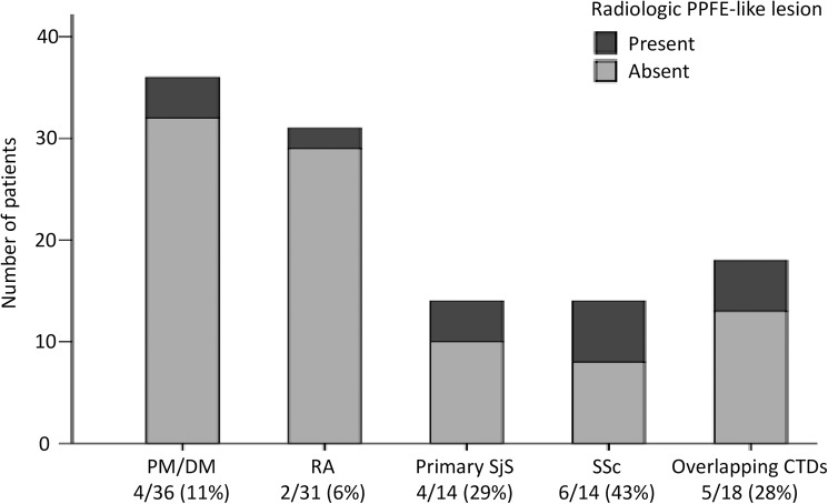
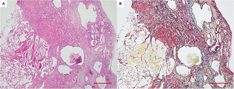
Radiologic PPFE-like lesion was found in 21 patients (19%) and were relatively frequent in those with systemic sclerosis (6/14: 43%) and primary Sjögren's syndrome (4/14: 29%). Patients with PPFE-like lesion were significantly older, had lower body mass index, higher ratio of residual volume to total lung capacity, and higher complication rate of pneumothorax and/or pneumomediastinum than those without. Twelve of the 21 patients were diagnosed radiologically as usual interstitial pneumonia (UIP) or possible UIP pattern. Two of three patients who underwent surgical lung biopsy of the upper lobes showed UIP on histopathology. Another patient was confirmed to have upper lobe PPFE on autopsy. During the clinical course, progression of the radiologic PPFE-like lesions was observed in 13 of 21 patients. Six patients died (mortality rate: 29%) and their PPFE-like lesions were commonly progressive. In the total cohort, our multivariate analysis identified the presence of PPFE-like lesion as a significant risk factor for respiratory death (hazard ratio: 4.10, 95% confidence interval: 1.33-12.65, p = 0.01).

CONCLUSION

In patients with CTD-related ILD, radiologic PPFE-like lesion, which may present as not only PPFE but also apical cap and upper lobe subpleural fibrosis commonly due to UIP, was not uncommon and was associated with poor prognosis. Clinicians should be cautious with this radiologic finding, particularly when it is progressive.

摘要

背景

在临床环境中偶尔可观察到包括肺尖帽在内的放射学胸膜实质纤维弹性组织增生症(PPFE)样病变。然而,在结缔组织病(CTD)相关的间质性肺疾病(ILD)中,放射学PPFE样病变的意义尚不清楚。

材料与方法

共纳入113例CTD相关ILD患者,评估其放射学PPFE样病变,该病变定义为胸部高分辨率计算机断层扫描显示双侧、上叶及胸膜下致密实变,伴或不伴胸膜增厚。对临床、放射学和病理学特征进行评估。

结果

21例患者(19%)发现有放射学PPFE样病变,在系统性硬化症患者(6/14:43%)和原发性干燥综合征患者(4/14:29%)中相对常见。与无PPFE样病变的患者相比,有PPFE样病变的患者年龄显著更大,体重指数更低,残气量与肺总量之比更高,气胸和/或纵隔气肿的并发症发生率更高。21例患者中有12例经放射学诊断为普通型间质性肺炎(UIP)或可能的UIP型。接受上叶手术肺活检的3例患者中有2例组织病理学显示为UIP。另一例患者尸检证实有上叶PPFE。在临床过程中,21例患者中有13例观察到放射学PPFE样病变进展。6例患者死亡(死亡率:29%),其PPFE样病变通常呈进行性。在整个队列中,我们的多变量分析确定PPFE样病变的存在是呼吸死亡的一个重要危险因素(风险比:4.10,95%置信区间:1.33 - 12.65,p = 0.01)。

结论

在CTD相关ILD患者中,放射学PPFE样病变不仅可表现为PPFE,还可表现为肺尖帽和通常由UIP引起的上叶胸膜下纤维化,并不罕见,且与预后不良相关。临床医生应对这一放射学表现保持谨慎,尤其是当其呈进行性时。

https://cdn.ncbi.nlm.nih.gov/pmc/blobs/8416/5493376/8c04596e3ea9/pone.0180283.g005.jpg
https://cdn.ncbi.nlm.nih.gov/pmc/blobs/8416/5493376/b757e9239487/pone.0180283.g001.jpg
https://cdn.ncbi.nlm.nih.gov/pmc/blobs/8416/5493376/6146cabc203e/pone.0180283.g002.jpg
https://cdn.ncbi.nlm.nih.gov/pmc/blobs/8416/5493376/0a61321efdbf/pone.0180283.g003.jpg
https://cdn.ncbi.nlm.nih.gov/pmc/blobs/8416/5493376/9f49445bca66/pone.0180283.g004.jpg
https://cdn.ncbi.nlm.nih.gov/pmc/blobs/8416/5493376/8c04596e3ea9/pone.0180283.g005.jpg

相似文献

1
Radiologic pleuroparenchymal fibroelastosis-like lesion in connective tissue disease-related interstitial lung disease.结缔组织病相关间质性肺疾病中的放射学胸膜实质纤维弹性组织增生样病变
PLoS One. 2017 Jun 30;12(6):e0180283. doi: 10.1371/journal.pone.0180283. eCollection 2017.
2
Clinical diagnosis of idiopathic pleuroparenchymal fibroelastosis: A retrospective multicenter study.特发性胸膜肺弹力纤维增生症的临床诊断:一项回顾性多中心研究。
Respir Med. 2017 Dec;133:1-5. doi: 10.1016/j.rmed.2017.11.003. Epub 2017 Nov 6.
3
Clinical significance of radiological pleuroparenchymal fibroelastosis pattern in interstitial lung disease patients registered for lung transplantation: a retrospective cohort study.影像学胸膜肺实质纤维弹性组织增生模式在登记肺移植的间质性肺病患者中的临床意义:一项回顾性队列研究。
Respir Res. 2018 Aug 30;19(1):162. doi: 10.1186/s12931-018-0860-6.
4
Lower-lobe predominant pleuroparenchymal fibroelastosis.下叶优势性胸膜肺弹力纤维增生症。
Pathol Int. 2019 Sep;69(9):536-540. doi: 10.1111/pin.12827. Epub 2019 Jul 4.
5
Pleuroparenchymal fibroelastosis in rheumatoid arthritis-associated interstitial lung disease.类风湿关节炎相关间质性肺疾病中的胸膜肺实质纤维弹性瘤。
Respir Res. 2022 Jun 2;23(1):143. doi: 10.1186/s12931-022-02064-z.
6
Pleuroparenchymal fibroelastosis in patients with idiopathic pulmonary fibrosis.特发性肺纤维化患者的胸膜肺实质纤维弹性化。
Respirology. 2020 Oct;25(10):1046-1052. doi: 10.1111/resp.13796. Epub 2020 Mar 9.
7
Distinct characteristics of pleuroparenchymal fibroelastosis with usual interstitial pneumonia compared with idiopathic pulmonary fibrosis.特发性肺纤维化与普通型间质性肺炎合并胸膜肺弹力纤维增生症的特征比较。
Chest. 2014 Nov;146(5):1248-1255. doi: 10.1378/chest.13-2866.
8
Various Bronchiolar Lesions Accompanied by Idiopathic Pleuroparenchymal Fibroelastosis with a Usual Interstitial Pneumonia Pattern Demonstrating Acute Exacerbation.伴有特发性胸膜肺纤维弹性组织增生症且呈普通间质性肺炎模式并显示急性加重的各种细支气管病变。
Intern Med. 2019 May 1;58(9):1321-1328. doi: 10.2169/internalmedicine.1649-18. Epub 2018 Dec 18.
9
Pleuroparenchymal fibroelastosis: a pattern of chronic lung injury.胸膜实质纤维弹性组织增生症:一种慢性肺损伤模式。
Hum Pathol. 2015 Jan;46(1):137-46. doi: 10.1016/j.humpath.2014.10.007. Epub 2014 Oct 17.
10
Pleuroparenchymal fibroelastosis as a histological background of autoimmune diseases.胸膜肺弹力纤维增生症作为自身免疫性疾病的组织学背景。
Virchows Arch. 2019 Jan;474(1):97-104. doi: 10.1007/s00428-018-2473-3. Epub 2018 Oct 16.

引用本文的文献

1
Korean Guidelines for the Diagnosis and Management of Interstitial Lung Disease: Other Forms of Interstitial Lung Disease.韩国间质性肺疾病诊断与管理指南:其他形式的间质性肺疾病
Tuberc Respir Dis (Seoul). 2025 Jul;88(3):454-476. doi: 10.4046/trd.2024.0181. Epub 2025 Mar 13.
2
The role of vascularity and the fibrovascular interface in interstitial lung diseases.血管形成及纤维血管界面在间质性肺疾病中的作用。
Eur Respir Rev. 2025 Feb 5;34(175). doi: 10.1183/16000617.0080-2024. Print 2025 Jan.
3
High resolution computed tomography in systemic sclerosis: From diagnosis to follow-up.

本文引用的文献

1
Pulmonary Apical Cap-What's Old Is New Again.肺尖帽——旧貌换新颜
Arch Pathol Lab Med. 2015 Oct;139(10):1258-62. doi: 10.5858/arpa.2015-0224-RA.
2
An official European Respiratory Society/American Thoracic Society research statement: interstitial pneumonia with autoimmune features.欧洲呼吸学会/美国胸科学会官方研究声明:具有自身免疫特征的间质性肺炎。
Eur Respir J. 2015 Oct;46(4):976-87. doi: 10.1183/13993003.00150-2015. Epub 2015 Jul 9.
3
Case records of the Massachusetts General Hospital. Case 14-2015. A 58-year-old woman with shortness of breath.
系统性硬化症的高分辨率计算机断层扫描:从诊断到随访
Rheumatol Immunol Res. 2024 Oct 21;5(3):166-174. doi: 10.2478/rir-2024-0023. eCollection 2024 Sep.
4
Proposed Clinical Algorithm for Pleuroparenchymal Fibroelastosis (PPFE).胸膜实质纤维弹性组织增生症(PPFE)的拟用临床算法
J Clin Med. 2024 Jun 24;13(13):3675. doi: 10.3390/jcm13133675.
5
Imaging in the diagnosis and management of fibrosing interstitial lung diseases.纤维化间质性肺疾病诊断与管理中的影像学检查
Breathe (Sheff). 2024 Mar;20(1):240006. doi: 10.1183/20734735.0006-2024. Epub 2024 May 14.
6
Multidisciplinary Approach to the Diagnosis of Idiopathic Interstitial Pneumonias: Focus on the Pathologist's Key Role.多学科方法诊断特发性间质性肺炎:关注病理学家的关键作用。
Int J Mol Sci. 2024 Mar 23;25(7):3618. doi: 10.3390/ijms25073618.
7
The role of lung biopsy for diagnosis and prognosis of interstitial lung disease in systemic sclerosis: a systematic literature review.系统性硬化症中肺活检对间质性肺疾病的诊断和预后的作用:系统文献回顾。
Respir Res. 2024 Mar 23;25(1):138. doi: 10.1186/s12931-024-02725-1.
8
Pulmonary fibroelastosis - A review.肺弹力纤维增生症——综述。
Matrix Biol. 2023 Dec;124:1-7. doi: 10.1016/j.matbio.2023.10.003. Epub 2023 Nov 3.
9
Pleuroparenchymal Fibroelastosis-like Lesions in Clinical Practice: A Rare Entity? Review of a Radiological Database.临床实践中的胸膜实质纤维弹性组织增生样病变:一种罕见实体?放射学数据库综述
Diagnostics (Basel). 2023 May 4;13(9):1627. doi: 10.3390/diagnostics13091627.
10
The world of rare interstitial lung diseases.罕见间质性肺疾病的世界。
Eur Respir Rev. 2023 Feb 7;32(167). doi: 10.1183/16000617.0161-2022. Print 2023 Mar 31.
马萨诸塞州总医院病例记录。病例14 - 2015。一名58岁呼吸急促的女性。
N Engl J Med. 2015 Apr 30;372(18):1749-58. doi: 10.1056/NEJMcpc1415200.
4
Pleuroparenchymal fibroelastosis from a consecutive database: a rare disease entity?来自连续数据库的胸膜实质纤维弹性组织增生症:一种罕见的疾病实体?
Eur Respir J. 2015 Apr;45(4):1183-6. doi: 10.1183/09031936.00214714. Epub 2015 Feb 19.
5
Distinct characteristics of pleuroparenchymal fibroelastosis with usual interstitial pneumonia compared with idiopathic pulmonary fibrosis.特发性肺纤维化与普通型间质性肺炎合并胸膜肺弹力纤维增生症的特征比较。
Chest. 2014 Nov;146(5):1248-1255. doi: 10.1378/chest.13-2866.
6
Predicting survival across chronic interstitial lung disease: the ILD-GAP model.预测慢性间质性肺病的生存情况:ILD-GAP 模型。
Chest. 2014 Apr;145(4):723-728. doi: 10.1378/chest.13-1474.
7
Prognostic factors in interstitial lung disease associated with primary Sjögren's syndrome: a retrospective analysis of 33 pathologically-proven cases.原发性干燥综合征相关间质性肺病的预后因素:33 例经病理证实病例的回顾性分析。
PLoS One. 2013 Sep 9;8(9):e73774. doi: 10.1371/journal.pone.0073774. eCollection 2013.
8
An official American Thoracic Society/European Respiratory Society statement: Update of the international multidisciplinary classification of the idiopathic interstitial pneumonias.美国胸科学会/欧洲呼吸学会官方声明:特发性间质性肺炎的国际多学科分类的更新。
Am J Respir Crit Care Med. 2013 Sep 15;188(6):733-48. doi: 10.1164/rccm.201308-1483ST.
9
Radiographic fibrosis score predicts survival in hypersensitivity pneumonitis.放射性纤维化评分可预测过敏性肺炎的生存情况。
Chest. 2013 Aug;144(2):586-592. doi: 10.1378/chest.12-2623.
10
Rapid decrease in forced vital capacity in patients with idiopathic pulmonary upper lobe fibrosis.特发性肺上叶纤维化患者用力肺活量的快速下降
Respir Investig. 2012 Sep;50(3):88-97. doi: 10.1016/j.resinv.2012.06.003. Epub 2012 Aug 1.